3
views
0
recommends
+1 Recommend
0 collections
    0
    shares
      • Record: found
      • Abstract: found
      • Article: found
      Is Open Access

      High‐density intracranial recordings reveal a distinct site in anterior dorsal precentral cortex that tracks perceived speech

      research-article

      Read this article at

      Bookmark
          There is no author summary for this article yet. Authors can add summaries to their articles on ScienceOpen to make them more accessible to a non-specialist audience.

          Abstract

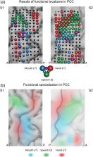
          Various brain regions are implicated in speech processing, and the specific function of some of them is better understood than others. In particular, involvement of the dorsal precentral cortex (dPCC) in speech perception remains debated, and attribution of the function of this region is more or less restricted to motor processing. In this study, we investigated high‐density intracranial responses to speech fragments of a feature film, aiming to determine whether dPCC is engaged in perception of continuous speech. Our findings show that dPCC exhibited preference to speech over other tested sounds. Moreover, the identified area was involved in tracking of speech auditory properties including speech spectral envelope, its rhythmic phrasal pattern and pitch contour. DPCC also showed the ability to filter out noise from the perceived speech. Comparing these results to data from motor experiments showed that the identified region had a distinct location in dPCC, anterior to the hand motor area and superior to the mouth articulator region. The present findings uncovered with high‐density intracranial recordings help elucidate the functional specialization of PCC and demonstrate the unique role of its anterior dorsal region in continuous speech perception.

          Abstract

          Berezutskaya et al. show that a distinct region within anterior dorsal precentral cortex tracks multiple auditory properties of perceived continuous speech. The region is distinct from the adjacent hand and mouth motor areas and has a unique role in speech processing.

          Related collections

          Most cited references101

          • Record: found
          • Abstract: found
          • Article: not found

          An automated labeling system for subdividing the human cerebral cortex on MRI scans into gyral based regions of interest.

          In this study, we have assessed the validity and reliability of an automated labeling system that we have developed for subdividing the human cerebral cortex on magnetic resonance images into gyral based regions of interest (ROIs). Using a dataset of 40 MRI scans we manually identified 34 cortical ROIs in each of the individual hemispheres. This information was then encoded in the form of an atlas that was utilized to automatically label ROIs. To examine the validity, as well as the intra- and inter-rater reliability of the automated system, we used both intraclass correlation coefficients (ICC), and a new method known as mean distance maps, to assess the degree of mismatch between the manual and the automated sets of ROIs. When compared with the manual ROIs, the automated ROIs were highly accurate, with an average ICC of 0.835 across all of the ROIs, and a mean distance error of less than 1 mm. Intra- and inter-rater comparisons yielded little to no difference between the sets of ROIs. These findings suggest that the automated method we have developed for subdividing the human cerebral cortex into standard gyral-based neuroanatomical regions is both anatomically valid and reliable. This method may be useful for both morphometric and functional studies of the cerebral cortex as well as for clinical investigations aimed at tracking the evolution of disease-induced changes over time, including clinical trials in which MRI-based measures are used to examine response to treatment.
            Bookmark
            • Record: found
            • Abstract: found
            • Article: not found

            FreeSurfer.

            FreeSurfer is a suite of tools for the analysis of neuroimaging data that provides an array of algorithms to quantify the functional, connectional and structural properties of the human brain. It has evolved from a package primarily aimed at generating surface representations of the cerebral cortex into one that automatically creates models of most macroscopically visible structures in the human brain given any reasonable T1-weighted input image. It is freely available, runs on a wide variety of hardware and software platforms, and is open source. Copyright © 2012 Elsevier Inc. All rights reserved.
              Bookmark
              • Record: found
              • Abstract: found
              • Article: not found

              The cortical organization of speech processing.

              Despite decades of research, the functional neuroanatomy of speech processing has been difficult to characterize. A major impediment to progress may have been the failure to consider task effects when mapping speech-related processing systems. We outline a dual-stream model of speech processing that remedies this situation. In this model, a ventral stream processes speech signals for comprehension, and a dorsal stream maps acoustic speech signals to frontal lobe articulatory networks. The model assumes that the ventral stream is largely bilaterally organized--although there are important computational differences between the left- and right-hemisphere systems--and that the dorsal stream is strongly left-hemisphere dominant.
                Bookmark

                Author and article information

                Contributors
                y.berezutskaya@umcutrecht.nl
                Journal
                Hum Brain Mapp
                Hum Brain Mapp
                10.1002/(ISSN)1097-0193
                HBM
                Human Brain Mapping
                John Wiley & Sons, Inc. (Hoboken, USA )
                1065-9471
                1097-0193
                03 August 2020
                November 2020
                : 41
                : 16 ( doiID: 10.1002/hbm.v41.16 )
                : 4587-4609
                Affiliations
                [ 1 ] Brain Center, Department of Neurology and Neurosurgery University Medical Center Utrecht Utrecht The Netherlands
                [ 2 ] Donders Institute for Brain, Cognition and Behaviour Radboud University Nijmegen The Netherlands
                [ 3 ] Université Grenoble Alpes Grenoble Institut des Neurosciences Grenoble France
                Author notes
                [*] [* ] Correspondence

                Julia Berezutskaya, Brain Center, Department of Neurology and Neurosurgery, University Medical Center Utrecht, Heidelberglaan 100, 3584 CX, Utrecht, The Netherlands.

                Email: y.berezutskaya@ 123456umcutrecht.nl

                Author information
                https://orcid.org/0000-0002-2005-8758
                Article
                HBM25144
                10.1002/hbm.25144
                7555065
                32744403
                8214a941-ab04-4fe4-8c45-ba139c07aba5
                © 2020 The Authors. Human Brain Mapping published by Wiley Periodicals LLC.

                This is an open access article under the terms of the http://creativecommons.org/licenses/by/4.0/ License, which permits use, distribution and reproduction in any medium, provided the original work is properly cited.

                History
                : 17 March 2020
                : 23 June 2020
                : 06 July 2020
                Page count
                Figures: 9, Tables: 2, Pages: 23, Words: 19443
                Funding
                Funded by: H2020 European Research Council , open-funder-registry 10.13039/100010663;
                Award ID: Advanced iConnect Project Grant ADV 320708
                Funded by: Nederlandse Organisatie voor Wetenschappelijk Onderzoek , open-funder-registry 10.13039/501100003246;
                Award ID: Language in Interaction Project Gravitation Grant
                Funded by: UMC Utrecht , open-funder-registry 10.13039/501100003761;
                Funded by: Netherlands Organisation for Scientific Research , open-funder-registry 10.13039/501100003246;
                Funded by: European Research Council , open-funder-registry 10.13039/100010663;
                Categories
                Research Article
                Research Articles
                Custom metadata
                2.0
                November 2020
                Converter:WILEY_ML3GV2_TO_JATSPMC version:5.9.2 mode:remove_FC converted:14.10.2020

                Neurology
                ecog,motor cortex,speech perception
                Neurology
                ecog, motor cortex, speech perception

                Comments

                Comment on this article