29
views
0
recommends
+1 Recommend
0 collections
    0
    shares
      • Record: found
      • Abstract: found
      • Article: found
      Is Open Access

      Age- and Brain Region-Specific Changes of Glucose Metabolic Disorder, Learning, and Memory Dysfunction in Early Alzheimer’s Disease Assessed in APP/PS1 Transgenic Mice Using 18F-FDG-PET

      research-article

      Read this article at

      Bookmark
          There is no author summary for this article yet. Authors can add summaries to their articles on ScienceOpen to make them more accessible to a non-specialist audience.

          Abstract

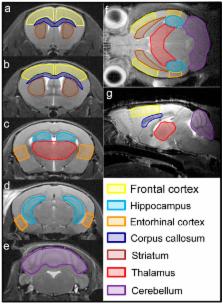
          Alzheimer’s disease (AD) is a leading cause of dementia worldwide, associated with cognitive deficits and brain glucose metabolic alteration. However, the associations of glucose metabolic changes with cognitive dysfunction are less detailed. Here, we examined the brains of APP/presenilin 1 (PS1) transgenic (Tg) mice aged 2, 3.5, 5 and 8 months using 18F-labed fluorodeoxyglucose ( 18F-FDG) microPET to assess age- and brain region-specific changes of glucose metabolism. FDG uptake was calculated as a relative standardized uptake value (SUVr). Morris water maze (MWM) was used to evaluate learning and memory dysfunction. We showed a glucose utilization increase in multiple brain regions of Tg mice at 2 and 3.5 months but not at 5 and 8 months. Comparisons of SUVrs within brains showed higher glucose utilization than controls in the entorhinal cortex, hippocampus, and frontal cortex of Tg mice at 2 and 3.5 months but in the thalamus and striatum at 3.5, 5 and 8 months. By comparing SUVrs in the entorhinal cortex and hippocampus, Tg mice were distinguished from controls at 2 and 3.5 months. In MWM, Tg mice aged 2 months shared a similar performance to the controls (prodromal-AD). By contrast, Tg mice failed training tests at 3.5 months but failed all MWM tests at 5 and 8 months, suggestive of partial or complete cognitive deficits (symptomatic-AD). Correlation analyses showed that hippocampal SUVrs were significantly correlated with MWM parameters in the symptomatic-AD stage. These data suggest that glucose metabolic disorder occurs before onset of AD signs in APP/PS1 mice with the entorhinal cortex and hippocampus affected first, and that regional FDG uptake increase can be an early biomarker for AD. Furthermore, hippocampal FDG uptake is a possible indicator for progression of Alzheimer’s cognition after cognitive decline, at least in animals.

          Related collections

          Most cited references34

          • Record: found
          • Abstract: found
          • Article: not found

          Discrimination between Alzheimer dementia and controls by automated analysis of multicenter FDG PET.

          A new diagnostic indicator of FDG PET scan abnormality, based on age-adjusted t statistics and an automated voxel-based procedure, is presented and validated in a large data set comprising 110 normal controls and 395 patients with probable Alzheimer's disease (AD) that were studied in eight participating centers. The effect of differences in spatial resolution of PET scanners was minimized effectively by filtering and masking. In controls FDG uptake declined significantly with age in anterior cingulate and frontolateral perisylvian cortex. In patients with probable AD decline of FDG uptake in posterior cingulate, temporoparietal, and prefrontal association cortex was related to dementia severity. These effects were clearly distinct from age effects in controls, suggesting that the disease process of AD is not related to normal aging. Women with probable AD had significantly more frontal metabolic impairment than men. The new indicator of metabolic abnormality in AD-related regions provided 93% sensitivity and specificity for distinction of mild to moderate probable AD from normals, and 84% sensitivity at 93% specificity for detection of very mild probable AD (defined by Mini Mental Score 24 or better). All regions related to AD severity were already affected in very mild AD, suggesting that all vulnerable areas are affected to a similar degree already at disease onset. Ventromedial frontal cortex was also abnormal. In conclusion, automated analysis of multicenter FDG PET is feasible, provides insights into AD pathophysiology, and can be used potentially as a sensitive biomarker for early AD diagnosis.
            Bookmark
            • Record: found
            • Abstract: found
            • Article: not found

            Comparing positron emission tomography imaging and cerebrospinal fluid measurements of β-amyloid.

            We examined agreement and disagreement between 2 biomarkers of β-amyloid (Aβ) deposition (amyloid positron emission tomography [PET] and cerebrospinal fluid [CSF] Aβ1-42 ) in normal aging and dementia in a large multicenter study. Concurrently acquired florbetapir PET and CSF Aβ were measured in cognitively normal, mild cognitive impairment (MCI), and Alzheimer's disease participants (n = 374) from the Alzheimer's Disease Neuroimaging Initiative. We also compared Aβ measurements in a separate group with serial CSF measurements over 3.1 ± 0.8 years that preceded a single florbetapir session. Additional biomarker and cognitive data allowed us to further examine profiles of discordant cases. Florbetapir and CSF Aβ were inversely correlated across all diagnostic groups, and dichotomous measurements were in agreement in 86% of subjects. Among subjects showing the most disagreement, the 2 discordant groups had different profiles: the florbetapir(+) /CSF Aβ(-) group was larger (n = 13) and was made up of only normal and early MCI subjects, whereas the florbetapir(-) /CSF Aβ(+) group was smaller (n = 7) and had poorer cognitive function and higher CSF tau, but no ApoE4 carriers. In the longitudinal sample, we observed both stable longitudinal CSF Aβ trajectories and those actively transitioning from normal to abnormal, but the final CSF Aβ measurements were in good agreement with florbetapir cortical retention. CSF and amyloid PET measurements of Aβ were consistent in the majority of subjects in the cross-sectional and longitudinal populations. Based on our analysis of discordant subjects, the available evidence did not show that CSF Aβ regularly becomes abnormal prior to fibrillar Aβ accumulation early in the course of disease. © 2013 American Neurological Association.
              Bookmark
              • Record: found
              • Abstract: found
              • Article: not found

              Glucose metabolism, gray matter structure, and memory decline in subjective memory impairment.

              To identify biological evidence for Alzheimer disease (AD) in individuals with subjective memory impairment (SMI) and unimpaired cognitive performance and to investigate the longitudinal cognitive course in these subjects. [¹⁸F]fluoro-2-deoxyglucose PET (FDG-PET) and structural MRI were acquired in 31 subjects with SMI and 56 controls. Cognitive follow-up testing was performed (average follow-up time: 35 months). Differences in baseline brain imaging data and in memory decline were assessed between both groups. Associations of memory decline with brain imaging data were tested. The SMI group showed hypometabolism in the right precuneus and hypermetabolism in the right medial temporal lobe. Gray matter volume was reduced in the right hippocampus in the SMI group. At follow-up, subjects with SMI showed a poorer performance than controls on measures of episodic memory. Longitudinal memory decline in the SMI group was associated with reduced glucose metabolism in the right precuneus at baseline. The cross-sectional difference in 2 independent neuroimaging modalities indicates early AD pathology in SMI. The poorer memory performance at follow-up and the association of reduced longitudinal memory performance with hypometabolism in the precuneus at baseline support the concept of SMI as the earliest manifestation of AD.
                Bookmark

                Author and article information

                Contributors
                Role: Academic Editor
                Journal
                Int J Mol Sci
                Int J Mol Sci
                ijms
                International Journal of Molecular Sciences
                MDPI
                1422-0067
                18 October 2016
                October 2016
                : 17
                : 10
                : 1707
                Affiliations
                [1 ]Department of Neurosurgery, Peking Union Medical College Hospital, Chinese Academy of Medical Sciences and Peking Union Medical College, Beijing 100730, China; lixueyuan-82@ 123456163.com (X.-Y.L.); zfx819_1986@ 123456163.com (F.-X.Z.)
                [2 ]Center for Magnetic Resonance Imaging, Peking University, Beijing 100871, China; wmen@ 123456pku.edu.cn
                [3 ]Department of Pathology, Comparative Medical Center, Peking Union Medical College & Institute of Laboratory Animal Science, Chinese Academy of Medical Science, Beijing 100021, China; zhuhua0226@ 123456vip.sina.com (H.Z.); wangjingzhan@ 123456sina.com (Z.-J.W.)
                [4 ]Center for Medical Experiments and Testing, Capital Medical University, Beijing 100069, China; ccmu2013@ 123456sina.com
                [5 ]Center for PET imaging, Peking Union Medical College Hospital, Chinese Academy of Medical Sciences and Peking Union Medical College, Beijing 100730, China; 13611093752@ 123456163.com
                Author notes
                [* ]Correspondence: baoxinjie1@ 123456pumch.cn (X.-J.B.); wangrz@ 123456126.com (R.-Z.W.); Tel./Fax: +86-10-6915-2532 (X.-J.B. & R.-Z.W.)
                Article
                ijms-17-01707
                10.3390/ijms17101707
                5085739
                27763550
                81ed00d8-bb77-4795-91e1-be410266529a
                © 2016 by the authors; licensee MDPI, Basel, Switzerland.

                This article is an open access article distributed under the terms and conditions of the Creative Commons Attribution (CC-BY) license ( http://creativecommons.org/licenses/by/4.0/).

                History
                : 31 August 2016
                : 04 October 2016
                Categories
                Article

                Molecular biology
                hippocampus,glucose metabolism,18f-fdg pet,cognitive dysfunction,app/ps1 mice,alzheimer’s disease

                Comments

                Comment on this article