32
views
0
recommends
+1 Recommend
0 collections
    0
    shares
      • Record: found
      • Abstract: found
      • Article: found
      Is Open Access

      Association between Elevated Pleural Interleukin-33 Levels and Tuberculous Pleurisy

      research-article

      Read this article at

      Bookmark
          There is no author summary for this article yet. Authors can add summaries to their articles on ScienceOpen to make them more accessible to a non-specialist audience.

          Abstract

          Background

          Interferon-γ (IFN-γ) plays a crucial role in Mycobacterium tuberculosis induced pleural responses. Interleukin (IL)-33 up-regulates the production of IFN-γ. We aimed to identify whether an association between pleural IL-33 levels and tuberculous pleurisy exists and determine its diagnostic value.

          Methods

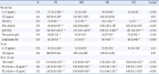
          Pleural IL-33, ST2 (a receptor of IL-33), adenosine deaminase (ADA), and IFN-γ, as well as serum IL-33 and ST2 were measured in 220 patients with pleural effusions (PEs). Patients with malignant (MPEs), parapneumonic (PPEs), tuberculous (TPEs), and cardiogenic (CPEs) pleural effusions were included.

          Results

          Pleural and serum IL-33 levels were highest or tended to be higher in patients with TPEs than in those with other types of PEs. The median pleural fluid-to-serum IL-33 ratio was higher in TPE cases (≥ 0.91) than in other PE cases (≤ 0.56). Pleural IL-33 levels correlated with those of pleural ADA and IFN-γ. However, the diagnostic accuracies of pleural IL-33 (0.74) and pleural fluid-to-serum IL-33 ratio (0.75) were lower than that of ADA (0.95) or IFN-γ (0.97). Pleural ST2 levels in patients with MPEs were higher than in patients with TPEs. Serum ST2 levels did not differ among the groups.

          Conclusions

          We identified an association between elevated pleural IL-33 levels and tuberculous pleurisy. However, we recommend conventional pleural markers (ADA or IFN-γ) as diagnostic markers of TPE.

          Related collections

          Most cited references30

          • Record: found
          • Abstract: found
          • Article: found
          Is Open Access

          Role of IL-33 in inflammation and disease

          Interleukin (IL)-33 is a new member of the IL-1 superfamily of cytokines that is expressed by mainly stromal cells, such as epithelial and endothelial cells, and its expression is upregulated following pro-inflammatory stimulation. IL-33 can function both as a traditional cytokine and as a nuclear factor regulating gene transcription. It is thought to function as an 'alarmin' released following cell necrosis to alerting the immune system to tissue damage or stress. It mediates its biological effects via interaction with the receptors ST2 (IL-1RL1) and IL-1 receptor accessory protein (IL-1RAcP), both of which are widely expressed, particularly by innate immune cells and T helper 2 (Th2) cells. IL-33 strongly induces Th2 cytokine production from these cells and can promote the pathogenesis of Th2-related disease such as asthma, atopic dermatitis and anaphylaxis. However, IL-33 has shown various protective effects in cardiovascular diseases such as atherosclerosis, obesity, type 2 diabetes and cardiac remodeling. Thus, the effects of IL-33 are either pro- or anti-inflammatory depending on the disease and the model. In this review the role of IL-33 in the inflammation of several disease pathologies will be discussed, with particular emphasis on recent advances.
            Bookmark
            • Record: found
            • Abstract: found
            • Article: not found

            Innate immunity to Mycobacterium tuberculosis.

            The different manifestations of infection with Mycobacterium tuberculosis reflect the balance between the bacillus and host defense mechanisms. Traditionally, protective immunity to tuberculosis has been ascribed to T-cell-mediated immunity, with CD4(+) T cells playing a crucial role. Recent immunological and genetic studies support the long-standing notion that innate immunity is also relevant in tuberculosis. In this review, emphasis is on these natural, innate host defense mechanisms, referring to experimental data (e.g., studies in gene knockout mice) and epidemiological, immunological, and genetic studies in human tuberculosis. The first step in the innate host defense is cellular uptake of M. tuberculosis, which involves different cellular receptors and humoral factors. Toll-like receptors seem to play a crucial role in immune recognition of M. tuberculosis, which is the next step. The subsequent inflammatory response is regulated by production of pro- and anti-inflammatory cytokines and chemokines. Different natural effector mechanisms for killing of M. tuberculosis have now been identified. Finally, the innate host response is necessary for induction of adaptive immunity to M. tuberculosis. These basic mechanisms augment our understanding of disease pathogenesis and clinical course and will be of help in designing adjunctive treatment strategies.
              Bookmark
              • Record: found
              • Abstract: found
              • Article: not found

              The pro-Th2 cytokine IL-33 directly interacts with invariant NKT and NK cells to induce IFN-gamma production.

              IL-33 has recently been identified as a cytokine endowed with pro-Th2 functions, raising the question of its effect on invariant natural killer T cell (iNKT), which are potent IL-4 producers. Here, we report a two-fold increase of iNKT-cell counts in spleen and liver after a 7-day treatment of mice with IL-33, which results from a direct effect, given that purified iNKT cells express the T1/ST2 receptor constitutively and respond to IL-33 by in vitro expansion and functional activation. Conversely to the expected pro-Th2 effect, IL-33 induced a preferential increase in IFN-gamma rather than IL-4 production upon TCR engagement that depended on endogenous IL-12. Moreover, in combination with the pro-inflammatory cytokine IL-12, IL-33 enhanced IFN-gamma production by both iNKT and NK cells. Taken together these data support the conclusion that IL-33 can contribute as a co-stimulatory factor to innate cellular immune responses.
                Bookmark

                Author and article information

                Journal
                Ann Lab Med
                Ann Lab Med
                ALM
                Annals of Laboratory Medicine
                The Korean Society for Laboratory Medicine
                2234-3806
                2234-3814
                January 2013
                17 December 2012
                : 33
                : 1
                : 45-51
                Affiliations
                [1 ]Department of Laboratory Medicine, Wonkwang University School of Medicine, Iksan, Korea.
                [2 ]Department of Internal Medicine, Wonkwang University School of Medicine, Iksan, Korea.
                [3 ]Department of Pathology, Wonkwang University School of Medicine, Iksan, Korea.
                [4 ]Department of Thoracic Surgery, Wonkwang University School of Medicine, Iksan, Korea.
                [5 ]Department of Otorhinolaryngology-Head and Neck Surgery, Wonkwang University School of Medicine, Iksan, Korea.
                [6 ]Department of Dermatology, Wonkwang University School of Medicine, Iksan, Korea.
                [7 ]Institute of Wonkwang Clinical Medicine & Genome Research Center for Immune Disorder, Wonkwang University School of Medicine, Iksan, Korea.
                [8 ]Institute of Wonkwang Medical Science & Center for Metabolic Function Regulation, Wonkwang University School of Medicine, Iksan, Korea.
                Author notes
                Corresponding author: Do-Sim Park. Department of Laboratory Medicine, Wonkwang University School of Medicine, 895 Muwang-ro, Iksan 570-711, Korea. Tel: +82-63-859-1863, Fax: +82-63-842-3786, emailds@ 123456hanmail.net

                *Koung-Sun Lee and Hak-Ryul Kim contributed equally to this work.

                Article
                10.3343/alm.2013.33.1.45
                3535196
                23301222
                81ba1d39-11ad-4468-8288-e1da99242511
                © The Korean Society for Laboratory Medicine.

                This is an Open Access article distributed under the terms of the Creative Commons Attribution Non-Commercial License ( http://creativecommons.org/licenses/by-nc/3.0/) which permits unrestricted non-commercial use, distribution, and reproduction in any medium, provided the original work is properly cited.

                History
                : 16 April 2012
                : 06 August 2012
                : 28 September 2012
                Categories
                Original Article
                Diagnostic Immunology

                Clinical chemistry
                interleukin-33,pleural effusion,st2,tuberculosis
                Clinical chemistry
                interleukin-33, pleural effusion, st2, tuberculosis

                Comments

                Comment on this article