28
views
0
recommends
+1 Recommend
0 collections
    0
    shares
      • Record: found
      • Abstract: found
      • Article: found
      Is Open Access

      The neurons that restore walking after paralysis

      research-article
      1 , 2 , 3 , 1 , 4 , 1 , 2 , 3 , 1 , 2 , 3 , 1 , 2 , 3 , 1 , 2 , 3 , 1 , 2 , 3 , 1 , 2 , 3 , 1 , 2 , 3 , 1 , 2 , 3 , 1 , 2 , 3 , 1 , 2 , 3 , 5 , 1 , 2 , 3 , 6 , 1 , 2 , 3 , 7 , 8 , 7 , 1 , 2 , 3 , 1 , 2 , 3 , 6 , 5 , 1 , 2 , 3 , 1 , 2 , 3 , , 1 , 2 , 3 , , 1 , 2 , 3 ,
      Nature
      Nature Publishing Group UK
      Spinal cord injury, Genetics of the nervous system

      Read this article at

      Bookmark
          There is no author summary for this article yet. Authors can add summaries to their articles on ScienceOpen to make them more accessible to a non-specialist audience.

          Abstract

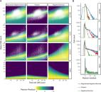
          A spinal cord injury interrupts pathways from the brain and brainstem that project to the lumbar spinal cord, leading to paralysis. Here we show that spatiotemporal epidural electrical stimulation (EES) of the lumbar spinal cord 13 applied during neurorehabilitation 4, 5 (EES REHAB) restored walking in nine individuals with chronic spinal cord injury. This recovery involved a reduction in neuronal activity in the lumbar spinal cord of humans during walking. We hypothesized that this unexpected reduction reflects activity-dependent selection of specific neuronal subpopulations that become essential for a patient to walk after spinal cord injury. To identify these putative neurons, we modelled the technological and therapeutic features underlying EES REHAB in mice. We applied single-nucleus RNA sequencing 69 and spatial transcriptomics 10, 11 to the spinal cords of these mice to chart a spatially resolved molecular atlas of recovery from paralysis. We then employed cell type 12, 13 and spatial prioritization to identify the neurons involved in the recovery of walking. A single population of excitatory interneurons nested within intermediate laminae emerged. Although these neurons are not required for walking before spinal cord injury, we demonstrate that they are essential for the recovery of walking with EES following spinal cord injury. Augmenting the activity of these neurons phenocopied the recovery of walking enabled by EES REHAB, whereas ablating them prevented the recovery of walking that occurs spontaneously after moderate spinal cord injury. We thus identified a recovery-organizing neuronal subpopulation that is necessary and sufficient to regain walking after paralysis. Moreover, our methodology establishes a framework for using molecular cartography to identify the neurons that produce complex behaviours.

          Abstract

          Transcriptomic analysis following epidural electrical stimulation of the lumbar spinal cord during neurorehabilitation in mice identifies a population of neurons that orchestrates the restoration of walking following paralysis.

          Related collections

          Most cited references84

          • Record: found
          • Abstract: found
          • Article: not found

          Comprehensive Integration of Single-Cell Data

          Single-cell transcriptomics has transformed our ability to characterize cell states, but deep biological understanding requires more than a taxonomic listing of clusters. As new methods arise to measure distinct cellular modalities, a key analytical challenge is to integrate these datasets to better understand cellular identity and function. Here, we develop a strategy to "anchor" diverse datasets together, enabling us to integrate single-cell measurements not only across scRNA-seq technologies, but also across different modalities. After demonstrating improvement over existing methods for integrating scRNA-seq data, we anchor scRNA-seq experiments with scATAC-seq to explore chromatin differences in closely related interneuron subsets and project protein expression measurements onto a bone marrow atlas to characterize lymphocyte populations. Lastly, we harmonize in situ gene expression and scRNA-seq datasets, allowing transcriptome-wide imputation of spatial gene expression patterns. Our work presents a strategy for the assembly of harmonized references and transfer of information across datasets.
            Bookmark
            • Record: found
            • Abstract: found
            • Article: found
            Is Open Access

            Normalization and variance stabilization of single-cell RNA-seq data using regularized negative binomial regression

            Single-cell RNA-seq (scRNA-seq) data exhibits significant cell-to-cell variation due to technical factors, including the number of molecules detected in each cell, which can confound biological heterogeneity with technical effects. To address this, we present a modeling framework for the normalization and variance stabilization of molecular count data from scRNA-seq experiments. We propose that the Pearson residuals from “regularized negative binomial regression,” where cellular sequencing depth is utilized as a covariate in a generalized linear model, successfully remove the influence of technical characteristics from downstream analyses while preserving biological heterogeneity. Importantly, we show that an unconstrained negative binomial model may overfit scRNA-seq data, and overcome this by pooling information across genes with similar abundances to obtain stable parameter estimates. Our procedure omits the need for heuristic steps including pseudocount addition or log-transformation and improves common downstream analytical tasks such as variable gene selection, dimensional reduction, and differential expression. Our approach can be applied to any UMI-based scRNA-seq dataset and is freely available as part of the R package sctransform, with a direct interface to our single-cell toolkit Seurat.
              Bookmark
              • Record: found
              • Abstract: found
              • Article: not found

              RNA velocity of single cells

              RNA abundance is a powerful indicator of the state of individual cells. Single-cell RNA sequencing can reveal RNA abundance with high quantitative accuracy, sensitivity and throughput1. However, this approach captures only a static snapshot at a point in time, posing a challenge for the analysis of time-resolved phenomena, such as embryogenesis or tissue regeneration. Here we show that RNA velocity—the time derivative of the gene expression state—can be directly estimated by distinguishing unspliced and spliced mRNAs in common single-cell RNA sequencing protocols. RNA velocity is a high-dimensional vector that predicts the future state of individual cells on a timescale of hours. We validate its accuracy in the neural crest lineage, demonstrate its use on multiple published datasets and technical platforms, reveal the branching lineage tree of the developing mouse hippocampus, and examine the kinetics of transcription in human embryonic brain. We expect RNA velocity to greatly aid the analysis of developmental lineages and cellular dynamics, particularly in humans.
                Bookmark

                Author and article information

                Contributors
                jocelyne.bloch@chuv.ch
                jordan.squair@epfl.ch
                gregoire.courtine@epfl.ch
                Journal
                Nature
                Nature
                Nature
                Nature Publishing Group UK (London )
                0028-0836
                1476-4687
                9 November 2022
                9 November 2022
                2022
                : 611
                : 7936
                : 540-547
                Affiliations
                [1 ]GRID grid.5333.6, ISNI 0000000121839049, Defitech Center for Interventional Neurotherapies (NeuroRestore), , EPFL/CHUV/UNIL, ; Lausanne, Switzerland
                [2 ]GRID grid.5333.6, ISNI 0000000121839049, NeuroX Institute and Brain Mind Institute, School of Life Sciences, , Swiss Federal Institute of Technology (EPFL), ; Lausanne, Switzerland
                [3 ]GRID grid.8515.9, ISNI 0000 0001 0423 4662, Department of Clinical Neuroscience, , Lausanne University Hospital (CHUV) and University of Lausanne (UNIL), ; Lausanne, Switzerland
                [4 ]GRID grid.17091.3e, ISNI 0000 0001 2288 9830, Michael Smith Laboratories, , University of British Columbia, ; Vancouver, British Columbia Canada
                [5 ]GRID grid.416870.c, ISNI 0000 0001 2177 357X, Spinal Circuits and Plasticity Unit, , National Institute of Neurological Disorders and Stroke, ; Bethesda, MD USA
                [6 ]GRID grid.5333.6, ISNI 0000000121839049, Bertarelli Foundation Chair in Neuroprosthetic Technology, Laboratory for Soft Bioelectronic Interfaces, , Institute of Electrical and Microengineering, Institute of Bioengineering, NeuroX Institute, EPFL, ; Geneva, Switzerland
                [7 ]GRID grid.8515.9, ISNI 0000 0001 0423 4662, Department of Nuclear Medicine and Molecular Imaging, , Lausanne University Hospital (CHUV) and University of Lausanne (UNIL), ; Lausanne, Switzerland
                [8 ]GRID grid.22937.3d, ISNI 0000 0000 9259 8492, Center for Medical Physics and Biomedical Engineering, , Medical University of Vienna, ; Vienna, Austria
                Author information
                http://orcid.org/0000-0001-6441-1755
                http://orcid.org/0000-0002-2168-1621
                http://orcid.org/0000-0001-6795-2688
                http://orcid.org/0000-0003-4910-8576
                http://orcid.org/0000-0003-0856-0929
                http://orcid.org/0000-0001-5233-8677
                http://orcid.org/0000-0001-7531-0036
                http://orcid.org/0000-0001-5018-5398
                http://orcid.org/0000-0003-1429-1374
                http://orcid.org/0000-0003-0894-1959
                http://orcid.org/0000-0001-9075-4022
                http://orcid.org/0000-0002-0335-0730
                http://orcid.org/0000-0002-9582-6109
                http://orcid.org/0000-0003-0551-859X
                http://orcid.org/0000-0002-5744-4142
                Article
                5385
                10.1038/s41586-022-05385-7
                9668750
                36352232
                7fe974dd-379a-47a5-8228-544a5da4cccc
                © The Author(s) 2022

                Open Access This article is licensed under a Creative Commons Attribution 4.0 International License, which permits use, sharing, adaptation, distribution and reproduction in any medium or format, as long as you give appropriate credit to the original author(s) and the source, provide a link to the Creative Commons license, and indicate if changes were made. The images or other third party material in this article are included in the article’s Creative Commons license, unless indicated otherwise in a credit line to the material. If material is not included in the article’s Creative Commons license and your intended use is not permitted by statutory regulation or exceeds the permitted use, you will need to obtain permission directly from the copyright holder. To view a copy of this license, visit http://creativecommons.org/licenses/by/4.0/.

                History
                : 9 November 2021
                : 23 September 2022
                Categories
                Article
                Custom metadata
                © The Author(s), under exclusive licence to Springer Nature Limited 2022

                Uncategorized
                spinal cord injury,genetics of the nervous system
                Uncategorized
                spinal cord injury, genetics of the nervous system

                Comments

                Comment on this article