99
views
0
recommends
+1 Recommend
0 collections
    0
    shares
      • Record: found
      • Abstract: found
      • Article: not found

      In Vivo Imaging of oskar mRNA Transport Reveals the Mechanism of Posterior Localization

      research-article

      Read this article at

      ScienceOpenPublisherPMC
      Bookmark
          There is no author summary for this article yet. Authors can add summaries to their articles on ScienceOpen to make them more accessible to a non-specialist audience.

          Summary

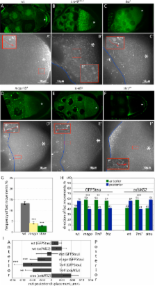
          oskar mRNA localization to the posterior of the Drosophila oocyte defines where the abdomen and germ cells form in the embryo. Although this localization requires microtubules and the plus end-directed motor, kinesin, its mechanism is controversial and has been proposed to involve active transport to the posterior, diffusion and trapping, or exclusion from the anterior and lateral cortex. By following oskar mRNA particles in living oocytes, we show that the mRNA is actively transported along microtubules in all directions, with a slight bias toward the posterior. This bias is sufficient to localize the mRNA and is reversed in mago, barentsz, and Tropomyosin II mutants, which mislocalize the mRNA anteriorly. Since almost all transport is mediated by kinesin, oskar mRNA localizes by a biased random walk along a weakly polarized cytoskeleton. We also show that each component of the oskar mRNA complex plays a distinct role in particle formation and transport.

          Related collections

          Most cited references52

          • Record: found
          • Abstract: found
          • Article: not found

          Single mRNA molecules demonstrate probabilistic movement in living mammalian cells.

          Cytoplasmic mRNA movements ultimately determine the spatial distribution of protein synthesis. Although some mRNAs are compartmentalized in cytoplasmic regions, most mRNAs, such as housekeeping mRNAs or the poly-adenylated mRNA population, are believed to be distributed throughout the cytoplasm. The general mechanism by which all mRNAs may move, and how this may be related to localization, is unknown. Here, we report a method to visualize single mRNA molecules in living mammalian cells, and we report that, regardless of any specific cytoplasmic distribution, individual mRNA molecules exhibit rapid and directional movements on microtubules. Importantly, the beta-actin mRNA zipcode increased both the frequency and length of these movements, providing a common mechanistic basis for both localized and nonlocalized mRNAs. Disruption of the cytoskeleton with drugs showed that microtubules and microfilaments are involved in the types of mRNA movements we have observed, which included complete immobility and corralled and nonrestricted diffusion. Individual mRNA molecules switched frequently among these movements, suggesting that mRNAs undergo continuous cycles of anchoring, diffusion, and active transport.
            Bookmark
            • Record: found
            • Abstract: found
            • Article: not found

            Oskar organizes the germ plasm and directs localization of the posterior determinant nanos.

            Oskar is one of seven Drosophila maternal-effect genes that are necessary for germline and abdomen formation. We have cloned oskar and show that oskar RNA is localized to the posterior pole of the oocyte when germ plasm forms. This polar distribution of oskar RNA is established during oogenesis in three phases: accumulation in the oocyte, transport toward the posterior, and finally maintenance at the posterior pole of the oocyte. The colocalization of oskar and nanos in wild-type and bicaudal embryos suggests that oskar directs localization of the posterior determinant nanos. We propose that the pole plasm is assembled stepwise and that continued interaction among its components is required for germ cell determination.
              Bookmark
              • Record: found
              • Abstract: found
              • Article: not found

              The autosomal FLP-DFS technique for generating germline mosaics in Drosophila melanogaster.

              The production of female germline chimeras is invaluable for analyzing the tissue specificity of recessive female sterile mutations as well as detecting the maternal effect of recessive zygotic lethal mutations. Previously, we developed the "FLP-DFS" technique to efficiently generate germline clones. This technique uses the X-linked germline-dependent dominant female sterile mutation ovoD1 as a selection for the detection of germline recombination events, and the FLP-FRT recombination system to promote site-specific chromosomal exchange. This method allows the efficient production of germline mosaics only on the X chromosome. In this paper we have built chromosomes that allow the use of this technique to the autosomes. We describe the various steps involved in the development of this technique as well as the properties of the chromosomes utilized.
                Bookmark

                Author and article information

                Journal
                Cell
                Cell
                Cell Press
                0092-8674
                1097-4172
                05 September 2008
                05 September 2008
                : 134
                : 5
                : 843-853
                Affiliations
                [1 ]The Gurdon Institute and the Department of Genetics, University of Cambridge, Tennis Court Road, CB2 1QN Cambridge, UK
                [2 ]Wellcome Trust Centre for Cell Biology, University of Edinburgh, Mayfield Road, Edinburgh EH9 3JR, UK
                Author notes
                []Corresponding author ds139@ 123456mole.bio.cam.ac.uk
                [3]

                Present address: Max Planck Institute of Molecular Cell Biology and Genetics, Pfotenhauerstr. 108, 01307 Dresden, Germany

                [4]

                Present address: The Gurdon Institute, University of Cambridge, Tennis Court Road, CB2 1QN Cambridge, UK

                [5]

                Present address: Department of Biochemistry, University of Oxford, Oxford OX1 3QU, UK

                [6]

                These authors contributed equally to this work

                Article
                CELL4040
                10.1016/j.cell.2008.06.053
                2585615
                18775316
                7fc7fe35-236e-4f9e-ad4d-f98974a2f6ee
                © 2008 ELL & Excerpta Medica.

                This document may be redistributed and reused, subject to certain conditions.

                History
                : 21 January 2008
                : 30 April 2008
                : 25 June 2008
                Categories
                Article

                Cell biology
                cellbio,devbio,rna
                Cell biology
                cellbio, devbio, rna

                Comments

                Comment on this article