57
views
0
recommends
+1 Recommend
0 collections
    0
    shares
      • Record: found
      • Abstract: found
      • Article: found
      Is Open Access

      Clinical genetics and pathobiology of ciliary chondrodysplasias

      research-article

      Read this article at

      Bookmark
          There is no author summary for this article yet. Authors can add summaries to their articles on ScienceOpen to make them more accessible to a non-specialist audience.

          Abstract

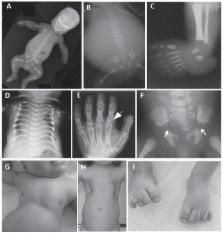
          Ciliary chondrodysplasias represent a heterogenous group of rare, nearly exclusively autosomal recessively inherited developmental conditions. While the skeletal phenotype, mainly affecting limbs, ribs and sometimes the craniofacial skeleton, is predominant, extraskeletal disease affecting the kidneys, liver, heart, eyes and other organs and tissues is observed inconsistently. Significant lethality, resulting from cardiorespiratory failure due to thoracic constriction as well as from renal and hepatic insufficiency or primary cardiac failure due to congenital heart disease, is observed with these conditions. The underlying genetic defects as well as developmental biology and cell biology work undertaken using animal model systems, suggest that these rare conditions result from ciliary malfunction. The skeletal phenotype is believed to result from imbalances in the hedgehog signaling pathway that normally occurs in functional cilia in chondrocytes. Although phenotypes have been historically distinguished based on clinical features into short-rib polydactyly syndrome, Jeune asphyxiating thoracic dystrophy, Mainzer-Saldino syndrome, Sensenbrenner syndrome (cranioectodermal dysplasia), oral-facial-digital syndrome and Ellis-van Creveld syndrome, recent research suggests that there is significant genetic as well as phenotypic overlap between the conditions. This review discusses ciliary chondrodysplasias from phenotypic hallmarks to clinical management and summarizes progress in identification of the underlying molecular mechanisms as well as potential future therapeutic perspectives.

          Related collections

          Most cited references104

          • Record: found
          • Abstract: found
          • Article: not found

          The vertebrate primary cilium in development, homeostasis, and disease.

          Cilia are complex structures that have garnered interest because of their roles in vertebrate development and their involvement in human genetic disorders. In contrast to multicellular invertebrates in which cilia are restricted to specific cell types, these organelles are found almost ubiquitously in vertebrate cells, where they serve a diverse set of signaling functions. Here, we highlight properties of vertebrate cilia, with particular emphasis on their relationship with other subcellular structures, and explore the physiological consequences of ciliary dysfunction.
            Bookmark
            • Record: found
            • Abstract: not found
            • Article: not found

            The primary cilium at a glance.

              Bookmark
              • Record: found
              • Abstract: found
              • Article: not found

              Inversin, the gene product mutated in nephronophthisis type II, functions as a molecular switch between Wnt signaling pathways.

              Cystic renal diseases are caused by mutations of proteins that share a unique subcellular localization: the primary cilium of tubular epithelial cells. Mutations of the ciliary protein inversin cause nephronophthisis type II, an autosomal recessive cystic kidney disease characterized by extensive renal cysts, situs inversus and renal failure. Here we report that inversin acts as a molecular switch between different Wnt signaling cascades. Inversin inhibits the canonical Wnt pathway by targeting cytoplasmic dishevelled (Dsh or Dvl1) for degradation; concomitantly, it is required for convergent extension movements in gastrulating Xenopus laevis embryos and elongation of animal cap explants, both regulated by noncanonical Wnt signaling. In zebrafish, the structurally related switch molecule diversin ameliorates renal cysts caused by the depletion of inversin, implying that an inhibition of canonical Wnt signaling is required for normal renal development. Fluid flow increases inversin levels in ciliated tubular epithelial cells and seems to regulate this crucial switch between Wnt signaling pathways during renal development.
                Bookmark

                Author and article information

                Journal
                101589859
                40543
                J Pediatr Genet
                J Pediatr Genet
                Journal of pediatric genetics
                2146-4596
                2146-460X
                6 December 2014
                November 2014
                11 December 2014
                : 3
                : 2
                : 46-94
                Affiliations
                [a ]Center for Pediatrics and Adolescent Medicine, University Hospital Freiburg, Freiburg, Germany
                [b ]Genetics and Genomics Medicine, Institute of Child Health, University College London, London, UK
                Author notes
                [* ]Corresponding author: Dr. Miriam Schmidts, MD, University Hospital Freiburg, Mathildenstrasse 1, 79112 Freiburg, Germany. Tel.: +44 791555 0317; miriam.schmidts@ 123456uniklinik-freiburg.de , miriam.schmidts.10@ 123456ucl.ac.uk
                Article
                EMS61288
                10.3233/PGE-14089
                4262788
                25506500
                7fb09e26-d2e5-4ea1-9c4f-3e7caae5c2a3
                © 2014 – IOS Press and the authors. All rights reserved

                This article is published online with Open Access and distributed under the terms of the Creative Commons Attribution Non-Commercial License.

                History
                Categories
                Article

                cilia,chondrodysplasia,jeune syndrome,short-rib polydactyly syndrome,sensenbrenner syndrome

                Comments

                Comment on this article