13
views
0
recommends
+1 Recommend
0 collections
    0
    shares
      • Record: found
      • Abstract: found
      • Article: found
      Is Open Access

      Ultrasonography Monitoring of Trauma-Induced Heterotopic Ossification: Guidance for Rehabilitation Procedures

      research-article

      Read this article at

      Bookmark
          There is no author summary for this article yet. Authors can add summaries to their articles on ScienceOpen to make them more accessible to a non-specialist audience.

          Abstract

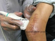
          Traumatic injury is one of varying causes of heterotopic ossification (HO). After HO occurrence, rehabilitation training need alterations to avoid the aggravation of HO. Therefore, monitoring of HO development plays an important role in the rehabilitation procedure. The aims of this study are to evaluate the post-traumatic HO occurring at various joints, to describe the features of HO development in ultrasound images, and to provide a guidance for the orthopedist to make individualized rehabilitation therapy. Eight subjects with the post-traumatic HO were recruited in this study. The joints on the injured side was examined by plain radiography. The joints on the injured side and the corresponding sites on the uninjured sides were scanned by ultrsonography. The HO tissues were segmented automatically using a semi-supervised segmentation algorithm. Then the HO tissues were evaluated in comparison with the corresponding region of the uninjured side. During the development stage of immature HO, ultrasonography was sensitive to observe the involved soft tissue and the calcification of HO. The characteristics of HO tissues in ultrasound image included the hyperechoic mass occasionally accompanied with acoustic shadow and the irregular muscular architecture. It was found that the mean grayscale value of HO was significantly higher ( p < 0.001) than that of the uninjured side at the middle and late stages. During the development period of HO, the HO grayscale value gradually increased and the mean grayscale of value of mature HO was significantly higher ( p < 0.05) than that of immature HO. According to the information of HO provided by ultrasound, the orthopedist properly adjusted the rehabilitation treatment. The results demonstrated that the visualization of HO using ultrasonography revealed the development of HO in the muscle tissues around the injured joints and thus provide a guidance for the orthopedist to make individualized rehabilitation therapy. Ultrasound could be a useful imaging modality for quantitative evaluation of HO during the rehabilitation of traumatic injury.

          Related collections

          Most cited references26

          • Record: found
          • Abstract: not found
          • Article: not found

          Ectopic ossification following total hip replacement. Incidence and a method of classification.

            Bookmark
            • Record: found
            • Abstract: found
            • Article: not found

            Skeletal muscle ultrasound: correlation between fibrous tissue and echo intensity.

            In this study, we examined the correlation between muscle ultrasound and muscle structure. Echo intensity (EI) of 14 muscles of two golden retriever muscular dystrophy dogs was correlated to the percentage interstitial fibrous tissue and fat in muscle biopsy. A significant correlation between interstitial fibrous tissue and EI was found (r = 0.87; p < 0.001). The separate influence of interstitial fat on muscle EI could not be established as only little fat was present. We conclude that fibrous tissue causes increased muscle EI. The high correlation between interstitial fibrous tissue and EI makes ultrasound a reliable method to determine severity of structural muscle changes.
              Bookmark
              • Record: found
              • Abstract: found
              • Article: not found

              Heterotopic ossification: a review.

              Heterotopic ossification is defined as the presence of lamellar bone at locations where bone normally does not exist. The condition must be distinguished from metastatic calcifications, which mainly occur in hypercalcaemia, and dystrophic calcifications in tumours. It is a frequent complication following central nervous system disorders (brain injuries, tumours, encephalitis, spinal cord lesions), multiple injuries, hip surgery and burns. In addition to this acquired form, hereditary causes also exist, such as fibrodysplasia ossificans progressiva, progressive osseous heteroplasia and Albright's hereditary osteodystrophy. Although these conditions are extremely rare, they can provide useful information on the physiopathology of heterotopic ossification, and thus lead to novel and causal treatment modalities. Heterotopic ossification is no trivial complication. A limitation of the range of joint motion may have serious consequences for the daily functioning of people who are already severely incapacitated because of their original lesion. Increased contractures and spasticity, pressure ulcers and increasing pain further compromise the patient's capabilities. Consequently, we feel that attention should be paid to the pathogenesis and particularly the prevention and treatment of this disorder.
                Bookmark

                Author and article information

                Contributors
                Journal
                Front Neurol
                Front Neurol
                Front. Neurol.
                Frontiers in Neurology
                Frontiers Media S.A.
                1664-2295
                13 September 2018
                2018
                : 9
                : 771
                Affiliations
                [1] 1Department of Biomedical Engineering, Southern Medical University , Guangzhou, China
                [2] 2Guangdong Work Injury Rehabilitation Center , Guangzhou, China
                [3] 3Guangdong Provincial Key Laboratory of Medical Image Processing, Southern Medical University , Guangzhou, China
                [4] 4Nanfang Hospital, Southern Medical University , Guangzhou, China
                Author notes

                Edited by: Xu Zhang, University of Science and Technology of China, China

                Reviewed by: Ke-Vin Chang, National Taiwan University Hospital, Taiwan; Wens Hou, Chongqing University, China

                *Correspondence: Qing Wang wq8740@ 123456smu.edu.cn

                This article was submitted to Neurotrauma, a section of the journal Frontiers in Neurology

                Article
                10.3389/fneur.2018.00771
                6149315
                30271377
                7f55c2c6-29eb-437d-8133-3d0544304a30
                Copyright © 2018 Wang, Zhang, Li, Song, Hu, Li, Chen and Wang.

                This is an open-access article distributed under the terms of the Creative Commons Attribution License (CC BY). The use, distribution or reproduction in other forums is permitted, provided the original author(s) and the copyright owner(s) are credited and that the original publication in this journal is cited, in accordance with accepted academic practice. No use, distribution or reproduction is permitted which does not comply with these terms.

                History
                : 30 June 2018
                : 24 August 2018
                Page count
                Figures: 6, Tables: 1, Equations: 0, References: 29, Pages: 9, Words: 5757
                Categories
                Neurology
                Original Research

                Neurology
                heterotopic ossification,ultrasonography,trauma,rehabilitation,diagnosis
                Neurology
                heterotopic ossification, ultrasonography, trauma, rehabilitation, diagnosis

                Comments

                Comment on this article