11
views
0
recommends
+1 Recommend
0 collections
    0
    shares
      • Record: found
      • Abstract: found
      • Article: found
      Is Open Access

      Altered Resting-State Functional Activity in Patients With Autism Spectrum Disorder: A Quantitative Meta-Analysis

      research-article

      Read this article at

      Bookmark
          There is no author summary for this article yet. Authors can add summaries to their articles on ScienceOpen to make them more accessible to a non-specialist audience.

          Abstract

          Background: There is accumulating evidence showing that patients with autism spectrum disorder (ASD) have obvious changes in resting-state functional brain activity. So far, there have been no meta-analyses of the resting-state brain activity alterations in patients with ASD. We attempted to explore the resting-state functional activity changes in patients with ASD, possibly providing a new perspective for investigating the pathophysiology of patients with ASD.

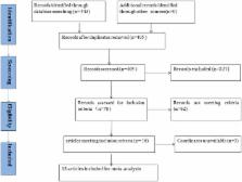
          Methods: We screened relevant studies published before August 2017 in PubMed, Ovid, Web of Science, China National Knowledge Infrastructure (CNKI), and the Wan-fang database. Fifteen resting-state functional neural activity datasets (including 382 patients and 348 healthy controls) were included. Through the use of the effect-size signed differential mapping (ES-SDM) method, we carried out a meta-analysis of resting-state functional activity studies of patients with ASD.

          Results: Compared with healthy controls, patients with ASD showed hyperactivity in the right supplementary motor area, middle frontal gyrus, inferior frontal gyrus, the left precentral gyrus, and the bilateral cerebellum hemispheric lobule (VIII/IX), and hypoactivity in the right middle temporal gyrus, superior temporal gyrus, and the left precuneus, posterior cingulate cortex, median cingulate cortex, and bilateral cerebellum (crus I).

          Conclusion: This meta-analysis indicates that patients with ASD have significant and robust resting-state brain activity alterations in the language comprehension network, inferior-posterior cerebellum, default mode network (DMN), and cerebellar crus I. These brain regions may serve as specific regions of interest for further studies of ASD, which will allow us to further clarify the neurobiological mechanisms in patients with ASD.

          Related collections

          Most cited references34

          • Record: found
          • Abstract: found
          • Article: not found

          Cortical activation and synchronization during sentence comprehension in high-functioning autism: evidence of underconnectivity.

          The brain activation of a group of high-functioning autistic participants was measured using functional MRI during sentence comprehension and the results compared with those of a Verbal IQ-matched control group. The groups differed in the distribution of activation in two of the key language areas. The autism group produced reliably more activation than the control group in Wernicke's (left laterosuperior temporal) area and reliably less activation than the control group in Broca's (left inferior frontal gyrus) area. Furthermore, the functional connectivity, i.e. the degree of synchronization or correlation of the time series of the activation, between the various participating cortical areas was consistently lower for the autistic than the control participants. These findings suggest that the neural basis of disordered language in autism entails a lower degree of information integration and synchronization across the large-scale cortical network for language processing. The article presents a theoretical account of the findings, related to neurobiological foundations of underconnectivity in autism.
            Bookmark
            • Record: found
            • Abstract: found
            • Article: not found

            Voxel-wise meta-analysis of grey matter changes in obsessive-compulsive disorder.

            Specific cortico-striato-thalamic circuits are hypothesised to mediate the symptoms of obsessive-compulsive disorder (OCD), but structural neuroimaging studies have been inconsistent. To conduct a meta-analysis of published and unpublished voxel-based morphometry studies in OCD. Twelve data-sets comprising 401 people with OCD and 376 healthy controls met inclusion criteria. A new improved voxel-based meta-analytic method, signed differential mapping (SDM), was developed to examine regions of increased and decreased grey matter volume in the OCD group v. control group. Results No between-group differences were found in global grey matter volumes. People with OCD had increased regional grey matter volumes in bilateral lenticular nuclei, extending to the caudate nuclei, as well as decreased volumes in bilateral dorsal medial frontal/anterior cingulate gyri. A descriptive analysis of quartiles, a sensitivity analysis as well as analyses of subgroups further confirmed these findings. Meta-regression analyses showed that studies that included individuals with more severe OCD were significantly more likely to report increased grey matter volumes in the basal ganglia. No effect of current antidepressant treatment was observed. Conclusions The results support a dorsal prefrontal-striatal model of the disorder and raise the question of whether functional alterations in other brain regions commonly associated with OCD, such as the orbitofrontal cortex, may reflect secondary compensatory strategies. Whether the reported differences between participants with OCD and controls precede the onset of the symptoms and whether they are specific to OCD remains to be established.
              Bookmark
              • Record: found
              • Abstract: found
              • Article: found
              Is Open Access

              The Neural Architecture of the Language Comprehension Network: Converging Evidence from Lesion and Connectivity Analyses

              While traditional models of language comprehension have focused on the left posterior temporal cortex as the neurological basis for language comprehension, lesion and functional imaging studies indicate the involvement of an extensive network of cortical regions. However, the full extent of this network and the white matter pathways that contribute to it remain to be characterized. In an earlier voxel-based lesion-symptom mapping analysis of data from aphasic patients (Dronkers et al., 2004), several brain regions in the left hemisphere were found to be critical for language comprehension: the left posterior middle temporal gyrus, the anterior part of Brodmann's area 22 in the superior temporal gyrus (anterior STG/BA22), the posterior superior temporal sulcus (STS) extending into Brodmann's area 39 (STS/BA39), the orbital part of the inferior frontal gyrus (BA47), and the middle frontal gyrus (BA46). Here, we investigated the white matter pathways associated with these regions using diffusion tensor imaging from healthy subjects. We also used resting-state functional magnetic resonance imaging data to assess the functional connectivity profiles of these regions. Fiber tractography and functional connectivity analyses indicated that the left MTG, anterior STG/BA22, STS/BA39, and BA47 are part of a richly interconnected network that extends to additional frontal, parietal, and temporal regions in the two hemispheres. The inferior occipito-frontal fasciculus, the arcuate fasciculus, and the middle and inferior longitudinal fasciculi, as well as transcallosal projections via the tapetum were found to be the most prominent white matter pathways bridging the regions important for language comprehension. The left MTG showed a particularly extensive structural and functional connectivity pattern which is consistent with the severity of the impairments associated with MTG lesions and which suggests a central role for this region in language comprehension.
                Bookmark

                Author and article information

                Contributors
                Journal
                Front Neurol
                Front Neurol
                Front. Neurol.
                Frontiers in Neurology
                Frontiers Media S.A.
                1664-2295
                24 July 2018
                2018
                : 9
                : 556
                Affiliations
                [1] 1Department of Radiology, Zhengzhou University People's Hospital and Henan Provincial People's Hospital , Zhengzhou, China
                [2] 2Department of Radiology, Union Hospital of Tongji Medical College, Huazhong University of Science and Technology , Wuhan, China
                [3] 3School of Life Science and Technology, Xidian University , Xi'an, China
                [4] 4Institute of Automation, Chinese Academy of Sciences , Beijing, China
                [5] 5Huaxi MR Research Center, Department of Radiology, West China Hospital of Sichuan University , Chengdu, China
                Author notes

                Edited by: Maria Assunta Rocca, San Raffaele Hospital (IRCCS), Italy

                Reviewed by: Martin Gorges, Universität Ulm, Germany; Domenico De Berardis, Azienda Usl Teramo, Italy

                *Correspondence: Meiyun Wang mywang@ 123456ha.edu.cn

                This article was submitted to Applied Neuroimaging, a section of the journal Frontiers in Neurology

                †These authors have contributed equally to this work.

                Article
                10.3389/fneur.2018.00556
                6066523
                29403429
                7f0844ff-ecff-4230-943a-8c213caee43a
                Copyright © 2018 Wang, Liu, Shi, Liu, Ma, Ma, Tian, Gong and Wang.

                This is an open-access article distributed under the terms of the Creative Commons Attribution License (CC BY). The use, distribution or reproduction in other forums is permitted, provided the original author(s) and the copyright owner(s) are credited and that the original publication in this journal is cited, in accordance with accepted academic practice. No use, distribution or reproduction is permitted which does not comply with these terms.

                History
                : 10 May 2017
                : 20 June 2018
                Page count
                Figures: 2, Tables: 3, Equations: 0, References: 53, Pages: 9, Words: 6166
                Funding
                Funded by: National Natural Science Foundation of China 10.13039/501100001809
                Award ID: 31470047
                Award ID: 81271565
                Categories
                Neurology
                Original Research

                Neurology
                autism spectrum disorder,meta-analysis,regional homogeneity,functional neuroimaging,resting state

                Comments

                Comment on this article