14
views
0
recommends
+1 Recommend
0 collections
    0
    shares
      • Record: found
      • Abstract: found
      • Article: found
      Is Open Access

      The chemokine receptor CCR10 promotes inflammation-driven hepatocarcinogenesis via PI3K/Akt pathway activation

      research-article

      Read this article at

      Bookmark
          There is no author summary for this article yet. Authors can add summaries to their articles on ScienceOpen to make them more accessible to a non-specialist audience.

          Abstract

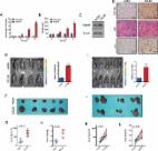
          G-protein-coupled receptor (GPCR)-related proteins are dysregulated and the GPCR CC-chemokine receptor 10 (CCR10) is significantly upregulated in inflammation-driven HCC. However, CCR10′s role in inflammation-driven hepatocarcinogenesis remains unknown. The aim of this study was to evaluate the role of CCR10 in inflammation-driven hepatocarcinogenesis. Via a targeted gene expression microarray screening alterations in GPCR family gene expression, we found CCR10 to be significantly upregulated in hepatocytes isolated from inflammation-driven human HCC tumors and matching paracancerous tissues. Tetrachloromethane (CCl4)-induced and diethylnitrosamine (DEN)-induced murine models of inflammatory hepatocarcinogenesis displayed significant hepatocellular TNF and CCR10 upregulation. Exogenous TNF applied to HepG2 and LO2 cell lines as well as wild-type (WT) mice significantly upregulated hepatocellular CCR10 expression, Akt phosphorylation, PCNA expression, and hepatocellular proliferation. Additionally, exogenous TNF significantly upregulated secretion of the natural CCR10 ligand-agonist CCL28 from both cell lines. Transgenic CCR10-knockout (CCR10 KO) in DEN-treated mice significantly increased hepatocellular apoptosis levels and significantly lowered compensatory hepatocellular proliferation but did not affect upstream TNF expression. In addition, DEN-treated CCR10 KO mice showed a significantly lower liver weight/body weight ratio, significantly lower liver tumor incidence, and significantly smaller tumors. Moreover, exogenous CCR10 expression significantly raised xenograft tumor growth in Balb/c nude mice. In vitro, CCR10 transfection or CCL28 treatment in HepG2 and LO2 cell lines significantly increased Akt phosphorylation, PCNA expression, and cell proliferation, while CCR10 silencing or Akt inhibition produced the opposite effects. In vivo, hepatocytes isolated from HCC tumor tissue and matching paracancerous tissue in DEN-treated CCR10 KO mice showed significantly lower Akt phosphorylation and PCNA expression relative to WT hepatocytes. In conclusion, inflammation-induced TNF promotes hepatocellular CCR10 expression and downstream PI3K/Akt-mediated hepatocarcinogenesis. CCR10 appears to function as a linkage between TNF stimulation and downstream PI3K/Akt pathway activation and shows promise as a potential therapeutic target for inflammation-driven HCC.

          Related collections

          Most cited references29

          • Record: found
          • Abstract: found
          • Article: not found
          Is Open Access

          Obesity, inflammation, and liver cancer.

          Obesity has become a universal and major public health problem with increasing prevalence in both adults and children in the 21st century, even in developing countries. Extensive epidemiological studies reveal a strong link between obesity and development and progression of various types of cancers. The connection between obesity and liver cancer is particularly strong and obesity often results in liver diseases such as non-alcoholic fatty liver disease (NAFLD) and the more severe non-alcoholic steatohepatitis (NASH). NASH is characterized by fatty liver inflammation and is believed to cause fibrosis and cirrhosis. The latter is a known liver cancer risk factor. In fact due to its much higher prevalence obesity may be a more substantial contributor to overall hepatocellular carcinoma burden than infection with hepatitis viruses. Here we review and discuss recent advances in elucidation of cellular and molecular alterations and signaling pathways associated with obesity and liver inflammation and their contribution to hepatocarcinogenesis. Copyright © 2011 European Association for the Study of the Liver. Published by Elsevier B.V. All rights reserved.
            Bookmark
            • Record: found
            • Abstract: found
            • Article: not found

            DCs metabolize sunlight-induced vitamin D3 to 'program' T cell attraction to the epidermal chemokine CCL27.

            During adaptive immune responses, dendritic cells activate T cells and endow them with specific homing properties. Mechanisms that 'imprint' specific tropisms, however, are not well defined. We show here that 1,25(OH)(2)D(3), the active form of vitamin D3, signaled T cells to express CC chemokine receptor 10, which enabled them to migrate to the skin-specific chemokine CCL27 secreted by keratinocytes of the epidermis. In contrast, 1,25(OH)(2)D(3) suppressed the gut-homing receptors alpha4beta7 and CCR9. Vitamin D3, the inactive prohormone naturally generated in the skin by exposure to the sun, was processed by dendritic cells and T cells to the active metabolite, providing a mechanism for the local regulation of T cell 'epidermotropism'. Our findings support a model in which dendritic cells process and 'interpret' locally produced metabolites to 'program' T cell homing and microenvironmental positioning.
              Bookmark
              • Record: found
              • Abstract: found
              • Article: found
              Is Open Access

              Hypoxia-induced CCL28 promotes recruitment of regulatory T cells and tumor growth in liver cancer

              Tumor cells craft microenvironment to overcome growth disadvantages and adjust to escape the immunosurveillance during tumorigenesis and metastasis. The evolving adaption to the changing microenvironment is exemplified by the development of strategies to deal with hypoxia resulted from fast proliferation of the tumor cells. In this study, we found that hypoxia hepatocellular carcinoma (HCC) cells recruited Regulatory T cells (Tregs) and expressed more Chemokine (C-C motif) ligand 28 (CCL28). Deletion of CCL28 inhibited Treg recruitment. Furthermore, overexpression of CCL28 promoted tumor growth and Treg infiltration in vivo. Enhanced angiogenesis and VEGF expression was also observed. Moreover, inhibition of HIF1α reversed hypoxia-induced CCL28 upregulation. Taken together, our results demonstrate that HCC recruits Tregs to promote angiogenesis under hypoxic condition by upregulating CCL28 expression. These findings establish a link between Tregs and hypoxia in HCC growth and may provide a new potential therapeutic target for treating HCC.
                Bookmark

                Author and article information

                Contributors
                (+86) 79186292163 , guowuhua@aliyun.com
                (+86) 79186268680 , jfzheng66@163.com
                Journal
                Cell Death Dis
                Cell Death Dis
                Cell Death & Disease
                Nature Publishing Group UK (London )
                2041-4889
                14 February 2018
                14 February 2018
                February 2018
                : 9
                : 2
                : 232
                Affiliations
                [1 ]GRID grid.412455.3, Department of Clinical Laboratory Medicine, , The Second Affiliated Hospital of Nanchang University, ; Nanchang, 330006 China
                [2 ]ISNI 0000 0004 0368 8293, GRID grid.16821.3c, Department of Gastrointestinal Surgery, Renji Hospital, , Shanghai Jiao Tong University School of Medicine, ; Shanghai, 200127 China
                [3 ]GRID grid.412455.3, Department of Gastroenterology, , The Second Affiliated Hospital of Nanchang University, ; Nanchang, 330006 China
                [4 ]GRID grid.443385.d, Department of Laboratory Medicine, , Affiliated Hospital of Guilin Medical University, ; Guilin, 541001 China
                Article
                267
                10.1038/s41419-018-0267-9
                5833857
                29445190
                7ec0e8e2-0674-493e-8cdf-c9c12c241cd5
                © The Author(s) 2018

                Open Access This article is licensed under a Creative Commons Attribution 4.0 International License, which permits use, sharing, adaptation, distribution and reproduction in any medium or format, as long as you give appropriate credit to the original author(s) and the source, provide a link to the Creative Commons license, and indicate if changes were made. The images or other third party material in this article are included in the article’s Creative Commons license, unless indicated otherwise in a credit line to the material. If material is not included in the article’s Creative Commons license and your intended use is not permitted by statutory regulation or exceeds the permitted use, you will need to obtain permission directly from the copyright holder. To view a copy of this license, visit http://creativecommons.org/licenses/by/4.0/.

                History
                : 20 October 2017
                : 27 November 2017
                : 28 November 2017
                Categories
                Article
                Custom metadata
                © The Author(s) 2018

                Cell biology
                Cell biology

                Comments

                Comment on this article