2
views
0
recommends
+1 Recommend
0 collections
    0
    shares
      • Record: found
      • Abstract: found
      • Article: found
      Is Open Access

      Crystal Structure of Refolding Fusion Core of Lassa Virus GP2 and Design of Lassa Virus Fusion Inhibitors

      research-article

      Read this article at

      Bookmark
          There is no author summary for this article yet. Authors can add summaries to their articles on ScienceOpen to make them more accessible to a non-specialist audience.

          Abstract

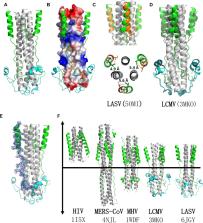
          The envelope glycoproteins GP1 and GP2 of Lassa virus (LASV) bind to the host cell receptors to mediate viral infection. So far, no approved vaccines and specific treatment options against LASV exist. To develop specific fusion inhibitors against LASV, we solved the crystal structure of the post-fusion 6 helix bundle (6-HB) formed by two heptad repeat domains (HR1 and HR2) of GP2. This fusion core contains a parallel trimeric coiled-coil of three HR1 helices, around which three HR2 helices are entwined in an antiparallel manner. Various hydrophobic and charged interactions form between HR1 and HR2 domains to stabilize the overall conformation of GP2 fusion core. Based on the structure, we designed several peptides spanning the HR2 domain and tested their antiviral activities. We found that the longer HR2 peptides were effective in inhibiting LASV GPC protein-mediated cell–cell fusion under low pH condition. These results not only suggest that LASV infects the target cell mainly through endocytosis, including micropinocytosis, and membrane fusion at low pH, but also provide an important basis for rational design of LASV fusion inhibitors.

          Related collections

          Most cited references18

          • Record: found
          • Abstract: found
          • Article: not found

          Automated structure solution with the PHENIX suite.

          Significant time and effort are often required to solve and complete a macromolecular crystal structure. The development of automated computational methods for the analysis, solution, and completion of crystallographic structures has the potential to produce minimally biased models in a short time without the need for manual intervention. The PHENIX software suite is a highly automated system for macromolecular structure determination that can rapidly arrive at an initial partial model of a structure without significant human intervention, given moderate resolution, and good quality data. This achievement has been made possible by the development of new algorithms for structure determination, maximum-likelihood molecular replacement (PHASER), heavy-atom search (HySS), template- and pattern-based automated model-building (RESOLVE, TEXTAL), automated macromolecular refinement (phenix. refine), and iterative model-building, density modification and refinement that can operate at moderate resolution (RESOLVE, AutoBuild). These algorithms are based on a highly integrated and comprehensive set of crystallographic libraries that have been built and made available to the community. The algorithms are tightly linked and made easily accessible to users through the PHENIX Wizards and the PHENIX GUI.
            Bookmark
            • Record: found
            • Abstract: found
            • Article: not found

            Identification of alpha-dystroglycan as a receptor for lymphocytic choriomeningitis virus and Lassa fever virus.

            A peripheral membrane protein that is interactive with lymphocytic choriomeningitis virus (LCMV) was purified from cells permissive to infection. Tryptic peptides from this protein were determined to be alpha-dystroglycan (alpha-DG). Several strains of LCMV and other arenaviruses, including Lassa fever virus (LFV), Oliveros, and Mobala, bound to purified alpha-DG protein. Soluble alpha-DG blocked both LCMV and LFV infection. Cells bearing a null mutation of the gene encoding DG were resistant to LCMV infection, and reconstitution of DG expression in null mutant cells restored susceptibility to LCMV infection. Thus, alpha-DG is a cellular receptor for both LCMV and LFV.
              Bookmark
              • Record: found
              • Abstract: found
              • Article: found
              Is Open Access

              Acidic pH-Induced Conformations and LAMP1 Binding of the Lassa Virus Glycoprotein Spike

              Lassa virus is an enveloped, bi-segmented RNA virus and the most prevalent and fatal of all Old World arenaviruses. Virus entry into the host cell is mediated by a tripartite surface spike complex, which is composed of two viral glycoprotein subunits, GP1 and GP2, and the stable signal peptide. Of these, GP1 binds to cellular receptors and GP2 catalyzes fusion between the viral envelope and the host cell membrane during endocytosis. The molecular structure of the spike and conformational rearrangements induced by low pH, prior to fusion, remain poorly understood. Here, we analyzed the three-dimensional ultrastructure of Lassa virus using electron cryotomography. Sub-tomogram averaging yielded a structure of the glycoprotein spike at 14-Å resolution. The spikes are trimeric, cover the virion envelope, and connect to the underlying matrix. Structural changes to the spike, following acidification, support a viral entry mechanism dependent on binding to the lysosome-resident receptor LAMP1 and further dissociation of the membrane-distal GP1 subunits.
                Bookmark

                Author and article information

                Contributors
                Journal
                Front Microbiol
                Front Microbiol
                Front. Microbiol.
                Frontiers in Microbiology
                Frontiers Media S.A.
                1664-302X
                13 August 2019
                2019
                : 10
                : 1829
                Affiliations
                [1] 1National Laboratory of Biomacromolecules, Institute of Biophysics, Chinese Academy of Sciences , Beijing, China
                [2] 2College of Life Sciences, University of Chinese Academy of Sciences , Beijing, China
                [3] 3Key Laboratory of Medical Molecular Virology of MOE/MOH/CAMS, School of Basic Medical Sciences and Shanghai Public Health Clinical Center, Fudan-Jinbo Joint Research Center, Fudan University , Shanghai, China
                [4] 4Interdisciplinary Innovation Institute of Medicine and Engineering, Beijing Advanced Innovation Center for Big Data-Based Precision Medicine, School of Biological Science and Medical Engineering, Beihang University , Beijing, China
                [5] 5Lindsley F. Kimball Research Institute, New York Blood Center , New York, NY, United States
                [6] 6National Center for Protein Science Shanghai, Shanghai Institute of Biochemistry and Cell Biology, Shanghai Institutes for Biological Sciences, Chinese Academy of Sciences , Shanghai, China
                Author notes

                Edited by: Lijun Rong, The University of Illinois at Chicago, United States

                Reviewed by: Jinsong Liu, Guangzhou Institutes of Biomedicine and Health (CAS), China; Yechiel Shai, Weizmann Institute of Science, Israel

                *Correspondence: Shibo Jiang, shibojiang@ 123456fudan.edu.cn
                Rongguang Zhang, rzhang@ 123456ibp.ac.cn

                These authors have contributed equally to this work

                This article was submitted to Virology, a section of the journal Frontiers in Microbiology

                Article
                10.3389/fmicb.2019.01829
                6700223
                31456769
                7e5e4f1d-1cf2-4e0a-b589-76068ffa3494
                Copyright © 2019 Zhang, Wang, Chen, Wang, Xu, Ye, Jiang, Zhu and Zhang.

                This is an open-access article distributed under the terms of the Creative Commons Attribution License (CC BY). The use, distribution or reproduction in other forums is permitted, provided the original author(s) and the copyright owner(s) are credited and that the original publication in this journal is cited, in accordance with accepted academic practice. No use, distribution or reproduction is permitted which does not comply with these terms.

                History
                : 04 June 2019
                : 25 July 2019
                Page count
                Figures: 6, Tables: 1, Equations: 0, References: 27, Pages: 12, Words: 0
                Categories
                Microbiology
                Original Research

                Microbiology & Virology
                lassa virus,gp2,fusion core,crystal structure,viral entry
                Microbiology & Virology
                lassa virus, gp2, fusion core, crystal structure, viral entry

                Comments

                Comment on this article