29
views
0
recommends
+1 Recommend
0 collections
    0
    shares
      • Record: found
      • Abstract: found
      • Article: found
      Is Open Access

      LPHN3, a presynaptic adhesion-GPCR implicated in ADHD, regulates the strength of neocortical layer 2/3 synaptic input to layer 5

      research-article

      Read this article at

      Bookmark
          There is no author summary for this article yet. Authors can add summaries to their articles on ScienceOpen to make them more accessible to a non-specialist audience.

          Abstract

          Background

          Latrophilins (LPHNs) are a small family of neuronal adhesion-GPCRs originally discovered as receptors for the black widow spider toxin α-latrotoxin. Mutations in LPHN3 have recently been identified as risk factors for attention deficit hyperactivity disorder (ADHD) in humans, but their physiological function has remained elusive. In this study, we tested two hypotheses regarding LPHN3 function: (1) LPHN3 regulates synaptic transmission by modulating probability of release; and (2) LPHN3 controls synapse development and the abundance of synapses.

          Results

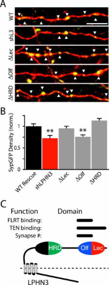
          We manipulated LPHN3 expression in mouse layer 2/3 (L2/3) pyramidal neurons and examined the consequences on the L2/3 to L5 cortical microcircuit. Employing an optogenetic strategy combined with shRNA knockdown of LPHN3, we found that LPHN3 did not influence probability of release at synapses formed by L2/3 neurons onto L5 pyramidal cells. The strength of L2/3 afferent input to L5, however, was weakened by loss of LPHN3. Using Synaptophysin-GFP as an anatomical marker of presynaptic terminals, we found that the density of synapses formed by L2/3 axons in L5 was reduced when LPHN3 was lost. Finally, we investigated the structural organization of the extracellular domain of LPHN3. We used single particle negative stain electron microscopy to image the extracellular domain of LPHN3 and showed that the Olfactomedin and Lectin domains form a globular domain on an elongated stalk. Cell-based binding experiments with mutant proteins revealed that the Olfactomedin domain was required for binding to FLRT3, whereas both the Olfactomedin and Lectin domains were involved in binding to Teneurin 1. Mutant LPHN3 lacking the Olfactomedin domain was not capable of rescuing the deficit in presynaptic density following knockdown of endogenous LPHN3.

          Conclusions

          We find that LPHN3 regulates the number of synapses formed by L2/3 neurons in L5 and the strength of synaptic drive from the L2/3-L5 pathway. The Olfactomedin domain of LPHN3 is required for this effect on synapse number and binding to its postsynaptic ligand FLRT3. We propose that LPHN3 functions in synaptic development and is important in determining the connectivity rates between principal neurons in the cortex.

          Related collections

          Most cited references22

          • Record: found
          • Abstract: found
          • Article: not found

          Negative Staining and Image Classification – Powerful Tools in Modern Electron Microscopy

          Vitrification is the state-of-the-art specimen preparation technique for molecular electron microscopy (EM) and therefore negative staining may appear to be an outdated approach. In this paper we illustrate the specific advantages of negative staining, ensuring that this technique will remain an important tool for the study of biological macromolecules. Due to the higher image contrast, much smaller molecules can be visualized by negative staining. Also, while molecules prepared by vitrification usually adopt random orientations in the amorphous ice layer, negative staining tends to induce preferred orientations of the molecules on the carbon support film. Combining negative staining with image classification techniques makes it possible to work with very heterogeneous molecule populations, which are difficult or even impossible to analyze using vitrified specimens.
            Bookmark
            • Record: found
            • Abstract: found
            • Article: found

            Latrophilin 1 and its endogenous ligand Lasso/teneurin-2 form a high-affinity transsynaptic receptor pair with signaling capabilities.

            Latrophilin 1 (LPH1), a neuronal receptor of α-latrotoxin, is implicated in neurotransmitter release and control of presynaptic Ca(2+). As an "adhesion G-protein-coupled receptor," LPH1 can convert cell surface interactions into intracellular signaling. To examine the physiological functions of LPH1, we used LPH1's extracellular domain to purify its endogenous ligand. A single protein of ∼275 kDa was isolated from rat brain and termed Lasso. Peptide sequencing and molecular cloning have shown that Lasso is a splice variant of teneurin-2, a brain-specific orphan cell surface receptor with a function in neuronal pathfinding and synaptogenesis. We show that LPH1 and Lasso interact strongly and specifically. They are always copurified from rat brain extracts. Coculturing cells expressing LPH1 with cells expressing Lasso leads to their mutual attraction and formation of multiple junctions to which both proteins are recruited. Cells expressing LPH1 form chimerical synapses with hippocampal neurons in cocultures; LPH1 and postsynaptic neuronal protein PSD-95 accumulate on opposite sides of these structures. Immunoblotting and immunoelectron microscopy of purified synapses and immunostaining of cultured hippocampal neurons show that LPH1 and Lasso are enriched in synapses; in both systems, LPH1 is presynaptic, whereas Lasso is postsynaptic. A C-terminal fragment of Lasso interacts with LPH1 and induces Ca(2+) signals in presynaptic boutons of hippocampal neurons and in neuroblastoma cells expressing LPH1. Thus, LPH1 and Lasso can form transsynaptic complexes capable of inducing presynaptic Ca(2+) signals, which might affect synaptic functions.
              Bookmark
              • Record: found
              • Abstract: found
              • Article: not found

              FLRT proteins are endogenous latrophilin ligands and regulate excitatory synapse development.

              Latrophilins (LPHNs) are a small family of G protein-coupled receptors known to mediate the massive synaptic exocytosis caused by the black widow spider venom α-latrotoxin, but their endogenous ligands and function remain unclear. Mutations in LPHN3 are strongly associated with attention deficit hyperactivity disorder, suggesting a role for latrophilins in human cognitive function. Using affinity chromatography and mass spectrometry, we identify the FLRT family of leucine-rich repeat transmembrane proteins as endogenous postsynaptic ligands for latrophilins. We demonstrate that the FLRT3 and LPHN3 ectodomains interact with high affinity in trans and that interference with this interaction using soluble recombinant LPHN3, LPHN3 shRNA, or FLRT3 shRNA reduces excitatory synapse density in cultured neurons. In addition, reducing FLRT3 levels with shRNA in vivo decreases afferent input strength and dendritic spine number in dentate granule cells. These observations indicate that LPHN3 and its ligand FLRT3 play an important role in glutamatergic synapse development. Copyright © 2012 Elsevier Inc. All rights reserved.
                Bookmark

                Author and article information

                Contributors
                Journal
                Neural Dev
                Neural Dev
                Neural Development
                BioMed Central
                1749-8104
                2014
                17 April 2014
                : 9
                : 7
                Affiliations
                [1 ]Neurobiology Section, Division of Biology, University of California San Diego, La Jolla, CA 92093, USA
                [2 ]Current affiliation: Duke University School of Medicine, Durham, NC 27710, USA
                [3 ]Child Health Institute of New Jersey and Department of Neuroscience and Cell Biology, Robert Wood Johnson Medical School, Rutgers University, New Brunswick, NJ 08901, USA
                [4 ]Neuroscience Discovery, F. Hoffmann-LaRoche, 4070 Basel, Switzerland
                Article
                1749-8104-9-7
                10.1186/1749-8104-9-7
                3996519
                24739570
                7e292525-d0a6-465c-91de-fd949f707040
                Copyright © 2014 O’Sullivan et al.; licensee BioMed Central Ltd.

                This is an Open Access article distributed under the terms of the Creative Commons Attribution License ( http://creativecommons.org/licenses/by/2.0), which permits unrestricted use, distribution, and reproduction in any medium, provided the original work is properly credited. The Creative Commons Public Domain Dedication waiver ( http://creativecommons.org/publicdomain/zero/1.0/) applies to the data made available in this article, unless otherwise stated.

                History
                : 29 January 2014
                : 31 March 2014
                Categories
                Research Article

                Neurosciences
                Neurosciences

                Comments

                Comment on this article