4
views
0
recommends
+1 Recommend
0 collections
    0
    shares
      • Record: found
      • Abstract: found
      • Article: found
      Is Open Access

      Effects of photobiomodulation on annulus fibrosus cells derived from degenerative disc disease patients exposed to microvascular endothelial cells conditioned medium

      research-article

      Read this article at

      Bookmark
          There is no author summary for this article yet. Authors can add summaries to their articles on ScienceOpen to make them more accessible to a non-specialist audience.

          Abstract

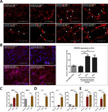
          Intervertebral disc (IVD) degeneration with chronic low back pain is associated with neo-vascularisation into the deeper IVD regions. During this process, endothelial cells (ECs), which are primarily responsible for angiogenesis, interact with the adjacent annulus fibrosus (AF) cells, which are the first line of defence against the invasion of vascular structures into deeper IVD regions. However, the accumulation of inflammatory and catabolic enzymes that results from this interaction promotes matrix degradation and an inflammatory response. Thus, regulating the production of these mediators and catabolic enzymes could ameliorate IVD degeneration. Photobiomodulation (PBM) therapy is a non-invasive stimulation known to have biologically beneficial effects on wound healing, tissue repair, and inflammation. Here, we examined the effects of PBM, administered at various wavelengths (645, 525, and 465 nm) and doses (16, 32, and 64 J/cm 2), on EC-stimulated human AF cells. Our results show that PBM selectively inhibited the EC-mediated production of inflammatory mediators, catabolic enzymes, and neurotrophins by human AF cells in a dose- and wavelength-dependent manner. These results suggest that PBM could be a superior and advanced treatment strategy for IVD degeneration.

          Related collections

          Most cited references32

          • Record: found
          • Abstract: found
          • Article: not found

          Role of cytokines in intervertebral disc degeneration: pain and disc content.

          Degeneration of the intervertebral discs (IVDs) is a major contributor to back, neck and radicular pain. IVD degeneration is characterized by increases in levels of the proinflammatory cytokines TNF, IL-1α, IL-1β, IL-6 and IL-17 secreted by the IVD cells; these cytokines promote extracellular matrix degradation, chemokine production and changes in IVD cell phenotype. The resulting imbalance in catabolic and anabolic responses leads to the degeneration of IVD tissues, as well as disc herniation and radicular pain. The release of chemokines from degenerating discs promotes the infiltration and activation of immune cells, further amplifying the inflammatory cascade. Leukocyte migration into the IVD is accompanied by the appearance of microvasculature tissue and nerve fibres. Furthermore, neurogenic factors, generated by both disc and immune cells, induce expression of pain-associated cation channels in the dorsal root ganglion. Depolarization of these ion channels is likely to promote discogenic and radicular pain, and reinforce the cytokine-mediated degenerative cascade. Taken together, an enhanced understanding of the contribution of cytokines and immune cells to these catabolic, angiogenic and nociceptive processes could provide new targets for the treatment of symptomatic disc disease. In this Review, the role of key inflammatory cytokines during each of the individual phases of degenerative disc disease, as well as the outcomes of major clinical studies aimed at blocking cytokine function, are discussed.
            Bookmark
            • Record: found
            • Abstract: found
            • Article: not found

            Intervertebral Disk Degeneration and Repair.

            Intervertebral disk (IVD) degeneration is a natural progression of the aging process. Degenerative disk disease (DDD) is a pathologic condition associated with IVD that has been associated with chronic back pain. There are a variety of different mechanisms of DDD (genetic, mechanical, exposure). Each of these pathways leads to a final common result of unbalancing the anabolic and catabolic environment of the extracellular matrix in favor of catabolism. Attempts have been made to gain an understanding of the process of IVD degeneration with in Vitro studies. These models help our understanding of the disease process, but are limited as they do not come close to replicating the complexities that exist with an in Vivo model. Animal models have been developed to help us gain further understanding of the degenerative cascade of IVD degeneration In Vivo and test experimental treatment modalities to either prevent or reverse the process of DDD. Many modalities for treatment of DDD have been developed including therapeutic protein injections, stem cell injections, gene therapy, and tissue engineering. These interventions have had promising outcomes in animal models. Several of these modalities have been attempted in human trials, with early outcomes having promising results. Further, increasing our understanding of the degenerative process is essential to the development of new therapeutic interventions and the optimization of existing treatment protocols. Despite limited data, biological therapies are a promising treatment modality for DDD that could impact our future management of low back pain.
              Bookmark
              • Record: found
              • Abstract: found
              • Article: not found

              Molecular mechanisms of biological aging in intervertebral discs.

              Advanced age is the greatest risk factor for the majority of human ailments, including spine-related chronic disability and back pain, which stem from age-associated intervertebral disc degeneration (IDD). Given the rapid global rise in the aging population, understanding the biology of intervertebral disc aging in order to develop effective therapeutic interventions to combat the adverse effects of aging on disc health is now imperative. Fortunately, recent advances in aging research have begun to shed light on the basic biological process of aging. Here we review some of these insights and organize the complex process of disc aging into three different phases to guide research efforts to understand the biology of disc aging. The objective of this review is to provide an overview of the current knowledge and the recent progress made to elucidate specific molecular mechanisms underlying disc aging. In particular, studies over the last few years have uncovered cellular senescence and genomic instability as important drivers of disc aging. Supporting evidence comes from DNA repair-deficient animal models that show increased disc cellular senescence and accelerated disc aging. Additionally, stress-induced senescent cells have now been well documented to secrete catabolic factors, which can negatively impact the physiology of neighboring cells and ECM. These along with other molecular drivers of aging are reviewed in depth to shed crucial insights into the underlying mechanisms of age-related disc degeneration. We also highlight molecular targets for novel therapies and emerging candidate therapeutics that may mitigate age-associated IDD. © 2016 Orthopaedic Research Society. Published by Wiley Periodicals, Inc. J Orthop Res 34:1289-1306, 2016.
                Bookmark

                Author and article information

                Contributors
                hyuk76@korea.ac.kr
                Journal
                Sci Rep
                Sci Rep
                Scientific Reports
                Nature Publishing Group UK (London )
                2045-2322
                15 June 2020
                15 June 2020
                2020
                : 10
                : 9655
                Affiliations
                [1 ]ISNI 0000 0001 0840 2678, GRID grid.222754.4, Department of Medical Sciences, Graduate School of Medicine, Korea University, ; Seoul, South Korea
                [2 ]Department of Neurosurgery, Guro Hospital, College of Medicine, Korea University, Seoul, Korea
                Article
                66689
                10.1038/s41598-020-66689-0
                7296027
                32541845
                7da7c798-1d2a-4335-88a2-11d1ef1d8beb
                © The Author(s) 2020

                Open Access This article is licensed under a Creative Commons Attribution 4.0 International License, which permits use, sharing, adaptation, distribution and reproduction in any medium or format, as long as you give appropriate credit to the original author(s) and the source, provide a link to the Creative Commons license, and indicate if changes were made. The images or other third party material in this article are included in the article’s Creative Commons license, unless indicated otherwise in a credit line to the material. If material is not included in the article’s Creative Commons license and your intended use is not permitted by statutory regulation or exceeds the permitted use, you will need to obtain permission directly from the copyright holder. To view a copy of this license, visit http://creativecommons.org/licenses/by/4.0/.

                History
                : 17 February 2020
                : 18 May 2020
                Categories
                Article
                Custom metadata
                © The Author(s) 2020

                Uncategorized
                cell biology,mechanisms of disease,biological techniques,optogenetics,biotechnology,tissue engineering,spine regulation and structure,chemokines,cytokines

                Comments

                Comment on this article