6
views
0
recommends
+1 Recommend
0 collections
    0
    shares
      • Record: found
      • Abstract: found
      • Article: found
      Is Open Access

      Omentin1 ameliorates myocardial ischemia-induced heart failure via SIRT3/FOXO3a-dependent mitochondrial dynamical homeostasis and mitophagy

      research-article

      Read this article at

      Bookmark
          There is no author summary for this article yet. Authors can add summaries to their articles on ScienceOpen to make them more accessible to a non-specialist audience.

          Abstract

          Background

          Adipose tissue-derived adipokines are involved in various crosstalk between adipose tissue and other organs. Omentin1, a novel adipokine, exerts vital roles in the maintenance of body metabolism, insulin resistance and the like. However, the protective effect of omentin1 in myocardial ischemia (MI)-induced heart failure (HF) and its specific mechanism remains unclear and to be elucidated.

          Methods

          The model of MI-induced HF mice and oxygen glucose deprivation (OGD)-injured cardiomyocytes were performed. Mice with overexpression of omentin1 were constructed by a fat-specific adeno-associated virus (AAV) vector system.

          Results

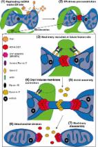
          We demonstrated that circulating omentin1 level diminished in HF patients compared with healthy subjects. Furthermore, the fat-specific overexpression of omentin1 ameliorated cardiac function, cardiac hypertrophy, infarct size and cardiac pathological features, and also enhanced SIRT3/FOXO3a signaling in HF mice. Additionally, administration with AAV-omentin1 increased mitochondrial fusion and decreased mitochondrial fission in HF mice, as evidenced by up-regulated expression of Mfn2 and OPA1, and downregulation of p-Drp1(Ser616). Then, it also promoted PINK1/Parkin-dependent mitophagy. Simultaneously, treatment with recombinant omentin1 strengthened OGD-injured cardiomyocyte viability, restrained LDH release, and enhanced the mitochondrial accumulation of SIRT3 and nucleus transduction of FOXO3a. Besides, omentin1 also ameliorated unbalanced mitochondrial fusion-fission dynamics and activated mitophagy, thereby, improving the damaged mitochondria morphology and controlling mitochondrial quality in OGD-injured cardiomyocytes. Interestingly, SIRT3 played an important role in the improvement effects of omentin1 on mitochondrial function, unbalanced mitochondrial fusion-fission dynamics and mitophagy.

          Conclusion

          Omentin1 improves MI-induced HF and myocardial injury by maintaining mitochondrial dynamical homeostasis and activating mitophagy via upregulation of SIRT3/FOXO3a signaling. This study provides evidence for further application of omentin1 in cardiovascular diseases from the perspective of crosstalk between heart and adipose tissue.

          Supplementary Information

          The online version contains supplementary material available at 10.1186/s12967-022-03642-x.

          Related collections

          Most cited references77

          • Record: found
          • Abstract: found
          • Article: not found

          A practical guide to evaluating colocalization in biological microscopy.

          Fluorescence microscopy is one of the most powerful tools for elucidating the cellular functions of proteins and other molecules. In many cases, the function of a molecule can be inferred from its association with specific intracellular compartments or molecular complexes, which is typically determined by comparing the distribution of a fluorescently labeled version of the molecule with that of a second, complementarily labeled probe. Although arguably the most common application of fluorescence microscopy in biomedical research, studies evaluating the "colocalization" of two probes are seldom quantified, despite a diversity of image analysis tools that have been specifically developed for that purpose. Here we provide a guide to analyzing colocalization in cell biology studies, emphasizing practical application of quantitative tools that are now widely available in commercial and free image analysis software.
            Bookmark
            • Record: found
            • Abstract: found
            • Article: found
            Is Open Access

            Mitochondrial dynamics: overview of molecular mechanisms

            Mitochondria are highly dynamic organelles undergoing coordinated cycles of fission and fusion, referred as ‘mitochondrial dynamics’, in order to maintain their shape, distribution and size. Their transient and rapid morphological adaptations are crucial for many cellular processes such as cell cycle, immunity, apoptosis and mitochondrial quality control. Mutations in the core machinery components and defects in mitochondrial dynamics have been associated with numerous human diseases. These dynamic transitions are mainly ensured by large GTPases belonging to the Dynamin family. Mitochondrial fission is a multi-step process allowing the division of one mitochondrion in two daughter mitochondria. It is regulated by the recruitment of the GTPase Dynamin-related protein 1 (Drp1) by adaptors at actin- and endoplasmic reticulum-mediated mitochondrial constriction sites. Drp1 oligomerization followed by mitochondrial constriction leads to the recruitment of Dynamin 2 to terminate membrane scission. Inner mitochondrial membrane constriction has been proposed to be an independent process regulated by calcium influx. Mitochondrial fusion is driven by a two-step process with the outer mitochondrial membrane fusion mediated by mitofusins 1 and 2 followed by inner membrane fusion, mediated by optic atrophy 1. In addition to the role of membrane lipid composition, several members of the machinery can undergo post-translational modifications modulating these processes. Understanding the molecular mechanisms controlling mitochondrial dynamics is crucial to decipher how mitochondrial shape meets the function and to increase the knowledge on the molecular basis of diseases associated with morphology defects. This article will describe an overview of the molecular mechanisms that govern mitochondrial fission and fusion in mammals.
              Bookmark
              • Record: found
              • Abstract: found
              • Article: found
              Is Open Access

              Adipose Tissue Dysfunction as Determinant of Obesity-Associated Metabolic Complications

              Obesity is a critical risk factor for the development of type 2 diabetes (T2D), and its prevalence is rising worldwide. White adipose tissue (WAT) has a crucial role in regulating systemic energy homeostasis. Adipose tissue expands by a combination of an increase in adipocyte size (hypertrophy) and number (hyperplasia). The recruitment and differentiation of adipose precursor cells in the subcutaneous adipose tissue (SAT), rather than merely inflating the cells, would be protective from the obesity-associated metabolic complications. In metabolically unhealthy obesity, the storage capacity of SAT, the largest WAT depot, is limited, and further caloric overload leads to the fat accumulation in ectopic tissues (e.g., liver, skeletal muscle, and heart) and in the visceral adipose depots, an event commonly defined as “lipotoxicity.” Excessive ectopic lipid accumulation leads to local inflammation and insulin resistance (IR). Indeed, overnutrition triggers uncontrolled inflammatory responses in WAT, leading to chronic low-grade inflammation, therefore fostering the progression of IR. This review summarizes the current knowledge on WAT dysfunction in obesity and its associated metabolic abnormalities, such as IR. A better understanding of the mechanisms regulating adipose tissue expansion in obesity is required for the development of future therapeutic approaches in obesity-associated metabolic complications.
                Bookmark

                Author and article information

                Contributors
                junpingkou@cpu.edu.cn
                lifang@cpu.edu.cn
                Journal
                J Transl Med
                J Transl Med
                Journal of Translational Medicine
                BioMed Central (London )
                1479-5876
                4 October 2022
                4 October 2022
                2022
                : 20
                : 447
                Affiliations
                [1 ]GRID grid.254147.1, ISNI 0000 0000 9776 7793, Jiangsu Key Laboratory of TCM Evaluation and Translational Research, Research Center for Traceability and Standardization of TCMs, School of Traditional Chinese Pharmacy, , China Pharmaceutical University, ; 639 Longmian Road, Nanjing, 211198 People’s Republic of China
                [2 ]GRID grid.410745.3, ISNI 0000 0004 1765 1045, Jiangsu Province Hospital of Chinese Medicine, , Affiliated Hospital of Nanjing University of Chinese Medicine, ; Nanjing, 210029 China
                Author information
                http://orcid.org/0000-0002-7617-6534
                Article
                3642
                10.1186/s12967-022-03642-x
                9531426
                36192726
                7d443d37-125b-492f-b697-fc759729a7d5
                © The Author(s) 2022

                Open AccessThis article is licensed under a Creative Commons Attribution 4.0 International License, which permits use, sharing, adaptation, distribution and reproduction in any medium or format, as long as you give appropriate credit to the original author(s) and the source, provide a link to the Creative Commons licence, and indicate if changes were made. The images or other third party material in this article are included in the article's Creative Commons licence, unless indicated otherwise in a credit line to the material. If material is not included in the article's Creative Commons licence and your intended use is not permitted by statutory regulation or exceeds the permitted use, you will need to obtain permission directly from the copyright holder. To view a copy of this licence, visit http://creativecommons.org/licenses/by/4.0/. The Creative Commons Public Domain Dedication waiver ( http://creativecommons.org/publicdomain/zero/1.0/) applies to the data made available in this article, unless otherwise stated in a credit line to the data.

                History
                : 22 July 2022
                : 16 September 2022
                Funding
                Funded by: FundRef http://dx.doi.org/10.13039/501100001809, National Natural Science Foundation of China;
                Award ID: 81973506
                Award Recipient :
                Categories
                Research
                Custom metadata
                © The Author(s) 2022

                Medicine
                omentin1,heart failure,sirtuin 3,mitochondrial dynamical homeostasis,mitophagy,heart-adipose crosstalk

                Comments

                Comment on this article