1
views
0
recommends
+1 Recommend
0 collections
    0
    shares
      • Record: found
      • Abstract: found
      • Article: found
      Is Open Access

      Ultra-high density electrodes improve detection, yield, and cell type specificity of brain recordings

      Preprint
      research-article

      Read this article at

      Bookmark
          There is no author summary for this article yet. Authors can add summaries to their articles on ScienceOpen to make them more accessible to a non-specialist audience.

          Abstract

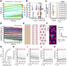
          To study the neural basis of behavior, we require methods to sensitively and accurately measure neural activity at single neuron and single spike resolution. Extracellular electrophysiology is a principal method for achieving this, but it has biases in the neurons it detects and it imperfectly resolves their action potentials. To overcome these limitations, we developed a silicon probe with significantly smaller and denser recording sites than previous designs, called Neuropixels Ultra (NP Ultra). This device measures neuronal activity at ultra-high densities (>1300 sites per mm, 10 times higher than previous probes), with 6 μm center-to-center spacing and low noise. This device effectively comprises an implantable voltage-sensing camera that captures a planar image of a neuron’s electrical field. We introduce a new spike sorting algorithm optimized for these probes and use it to find that the yield of visually-responsive neurons in recordings from mouse visual cortex improves ~3-fold. Recordings across multiple brain regions and four species revealed a subset of unexpectedly small extracellular action potentials not previously reported. Further experiments determined that, in visual cortex, these do not correspond to major subclasses of interneurons and instead likely reflect recordings from axons. Finally, using ground-truth identification of cortical inhibitory cell types with optotagging, we found that cell type was discriminable with approximately 75% success among three types, a significant improvement over lower-resolution recordings. NP Ultra improves spike sorting performance, sampling bias, and cell type classification.

          Related collections

          Most cited references63

          • Record: found
          • Abstract: found
          • Article: not found

          The origin of extracellular fields and currents--EEG, ECoG, LFP and spikes.

          Neuronal activity in the brain gives rise to transmembrane currents that can be measured in the extracellular medium. Although the major contributor of the extracellular signal is the synaptic transmembrane current, other sources--including Na(+) and Ca(2+) spikes, ionic fluxes through voltage- and ligand-gated channels, and intrinsic membrane oscillations--can substantially shape the extracellular field. High-density recordings of field activity in animals and subdural grid recordings in humans, combined with recently developed data processing tools and computational modelling, can provide insight into the cooperative behaviour of neurons, their average synaptic input and their spiking output, and can increase our understanding of how these processes contribute to the extracellular signal.
            Bookmark
            • Record: found
            • Abstract: found
            • Article: not found

            Fully integrated silicon probes for high-density recording of neural activity

            Sensory, motor and cognitive operations involve the coordinated action of large neuronal populations across multiple brain regions in both superficial and deep structures. Existing extracellular probes record neural activity with excellent spatial and temporal (sub-millisecond) resolution, but from only a few dozen neurons per shank. Optical Ca2+ imaging offers more coverage but lacks the temporal resolution needed to distinguish individual spikes reliably and does not measure local field potentials. Until now, no technology compatible with use in unrestrained animals has combined high spatiotemporal resolution with large volume coverage. Here we design, fabricate and test a new silicon probe known as Neuropixels to meet this need. Each probe has 384 recording channels that can programmably address 960 complementary metal–oxide–semiconductor (CMOS) processing-compatible low-impedance TiN sites that tile a single 10-mm long, 70 × 20-μm cross-section shank. The 6 × 9-mm probe base is fabricated with the shank on a single chip. Voltage signals are filtered, amplified, multiplexed and digitized on the base, allowing the direct transmission of noise-free digital data from the probe. The combination of dense recording sites and high channel count yielded well-isolated spiking activity from hundreds of neurons per probe implanted in mice and rats. Using two probes, more than 700 well-isolated single neurons were recorded simultaneously from five brain structures in an awake mouse. The fully integrated functionality and small size of Neuropixels probes allowed large populations of neurons from several brain structures to be recorded in freely moving animals. This combination of high-performance electrode technology and scalable chip fabrication methods opens a path towards recording of brain-wide neural activity during behaviour.
              Bookmark
              • Record: found
              • Abstract: found
              • Article: not found

              Spontaneous behaviors drive multidimensional, brainwide activity

              Neuronal populations in sensory cortex produce variable responses to sensory stimuli and exhibit intricate spontaneous activity even without external sensory input. Cortical variability and spontaneous activity have been variously proposed to represent random noise, recall of prior experience, or encoding of ongoing behavioral and cognitive variables. Recording more than 10,000 neurons in mouse visual cortex, we observed that spontaneous activity reliably encoded a high-dimensional latent state, which was partially related to the mouse’s ongoing behavior and was represented not just in visual cortex but also across the forebrain. Sensory inputs did not interrupt this ongoing signal but added onto it a representation of external stimuli in orthogonal dimensions. Thus, visual cortical population activity, despite its apparently noisy structure, reliably encodes an orthogonal fusion of sensory and multidimensional behavioral information.
                Bookmark

                Author and article information

                Journal
                bioRxiv
                BIORXIV
                bioRxiv
                Cold Spring Harbor Laboratory
                25 August 2023
                : 2023.08.23.554527
                Affiliations
                [1 ]Department of Biological Structure, University of Washington, Seattle, WA, USA
                [2 ]MindScope Program, Allen Institute, Seattle, WA, USA
                [3 ]Allen Institute for Neural Dynamics, Seattle, WA, USA
                [4 ]Department of Neuroscience, Zuckerman Mind Brain Behavior Institute, Columbia University, New York, NY, USA
                [5 ]Janelia Research Campus, Ashburn, VA, USA
                [6 ]Washington National Primate Research Center, Seattle, WA, USA
                [7 ]Max Planck Institute for Brain Research, Frankfurt, Germany
                [8 ]IMEC, Leuven, Belgium
                [9 ]Norwegian University of Life Sciences, Ås, Norway
                [10 ]University of Oslo, Oslo, Norway
                [11 ]Biomedical Engineering Department, Johns Hopkins University, Baltimore, MD, USA
                Author notes
                [# ]Correspondence to nick.steinmetz@ 123456gmail.com
                [*]

                equal contribution

                Author contributions

                ZY collected brain-wide acute mouse recordings. AMS and SM collected data with opto-tagging. ZY and JRS collected data with imposed probe motion and visual stimuli. JC performed noise and gain measurements. AMS performed light sensitivity measurements. SC collected acute mouse recordings. JHS wrote software for data acquisition. TN and WB collected non-human primate recordings. FP and NBS collected recordings from electric fish. SW collected recordings from bearded dragon lizards. JB, CW, CH, and LP developed the spike sorting methods. ZY, AMS, JC, JB, JRS, and NAS analyzed data. CML, BR, and BD performed engineering and fabrication of NP Ultra probes. TVN wrote the computational model and NAS analyzed the output. ZY, AMS, JC, JB, JRS, LP, SRO, and NAS wrote the original draft manuscript. All authors reviewed and edited the manuscript. SRO, TDH, and NAS obtained primary funding. GTE, GL, NBS, WB, AP, CML, BD, LP, JHS, CK, SRO, TDH, and NAS supervised aspects of the work.

                Article
                10.1101/2023.08.23.554527
                10473688
                37662298
                7d338c70-b64f-4ec5-bf2a-00e6c801209f

                This work is licensed under a Creative Commons Attribution-NonCommercial 4.0 International License, which allows reusers to distribute, remix, adapt, and build upon the material in any medium or format for noncommercial purposes only, and only so long as attribution is given to the creator.

                History
                Funding
                Funded by: NIH BRAIN Initiative, NAS, SRO, TDH
                Award ID: U01NS113252
                Funded by: Pew Biomedical Scholars Program, NAS, Klingenstein-Simons Fellowship in Neuroscience, NAS, Max Planck Society, GL, European Research Council, European Union’s Horizon 2020 research and innovation programme, GL, NIH, NBS, NSF, NBS
                Award ID: 834446, R01 NS118448, R01 NS075023, IOS 211500
                Funded by: NIH, WaNPRC, NIH
                Award ID: P51 (OD010425), U42 (OD011123)
                Funded by: European Union Horizon 2020 Research and Innovation Programme, Human Brain Project SGA3, GTE, TVN
                Award ID: 945539
                Categories
                Article

                Comments

                Comment on this article