51
views
0
recommends
+1 Recommend
1 collections
    0
    shares
      • Record: found
      • Abstract: found
      • Article: found
      Is Open Access

      Structural brain abnormalities in the common epilepsies assessed in a worldwide ENIGMA study

      research-article
      1 , 2 , 3 , 4 , 1 , 1 , 5 , 2 , 6 , 7 , 8 , 9 , 10 , 11 , 7 , 7 , 12 , 13 , 7 , 14 , 15 , 16 , 17 , 18 , 19 , 14 , 5 , 20 , 21 , 7 , 22 , 23 , 1 , 24 , 25 , 26 , 27 , 12 , 20 , 10 , 28 , 29 , 30 , 28 , 29 , 25 , 31 , 32 , 7 , 15 , 33 , 34 , 9 , 35 , 27 , 36 , 37 , 38 , 39 , 36 , 39 , 40 , 41 , 42 , 43 , 44 , 43 , 45 , 46 , 7 , 47 , 15 , 48 , 26 , 30 , 30 , 30 , 49 , 23 , 12 , 50 , 2 , 51 , 7 , 33 , 2 , 51 , 52 , 53 , 12 , 51 , 54 , 25 , 55 , 14 , 56 , 10 , 11 , 37 , 38 , 27 , 57 , 8 , 9 , 58 , 41 , 14 , 56 , 28 , 29 , 39 , 41 , 59 , 3 , 16 , 60 , 61 , 12 , 13 , 34 , 18 , 19 , 62 , 4 , 63 , 1 , 64 , 65 ,
      Brain
      Oxford University Press
      epilepsy, MRI, thalamus, precentral gyrus

      Read this article at

      ScienceOpenPublisherPMC
      Bookmark
          There is no author summary for this article yet. Authors can add summaries to their articles on ScienceOpen to make them more accessible to a non-specialist audience.

          Abstract

          Structural MRI abnormalities are inconsistently reported in epilepsy. In the largest neuroimaging study to date, Whelan et al. report robust structural alterations across and within epilepsy syndromes, including shared volume loss in the thalamus, and widespread cortical thickness differences. The resulting neuroanatomical map will guide prospective studies of disease progression.

          Abstract

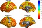
          Progressive functional decline in the epilepsies is largely unexplained. We formed the ENIGMA-Epilepsy consortium to understand factors that influence brain measures in epilepsy, pooling data from 24 research centres in 14 countries across Europe, North and South America, Asia, and Australia. Structural brain measures were extracted from MRI brain scans across 2149 individuals with epilepsy, divided into four epilepsy subgroups including idiopathic generalized epilepsies ( n =367), mesial temporal lobe epilepsies with hippocampal sclerosis (MTLE; left, n = 415; right, n = 339), and all other epilepsies in aggregate ( n = 1026), and compared to 1727 matched healthy controls. We ranked brain structures in order of greatest differences between patients and controls, by meta-analysing effect sizes across 16 subcortical and 68 cortical brain regions. We also tested effects of duration of disease, age at onset, and age-by-diagnosis interactions on structural measures. We observed widespread patterns of altered subcortical volume and reduced cortical grey matter thickness. Compared to controls, all epilepsy groups showed lower volume in the right thalamus (Cohen’s d = −0.24 to −0.73; P < 1.49 × 10 −4), and lower thickness in the precentral gyri bilaterally ( d = −0.34 to −0.52; P < 4.31 × 10 −6). Both MTLE subgroups showed profound volume reduction in the ipsilateral hippocampus ( d = −1.73 to −1.91, P < 1.4 × 10 −19), and lower thickness in extrahippocampal cortical regions, including the precentral and paracentral gyri, compared to controls ( d = −0.36 to −0.52; P < 1.49 × 10 −4). Thickness differences of the ipsilateral temporopolar, parahippocampal, entorhinal, and fusiform gyri, contralateral pars triangularis, and bilateral precuneus, superior frontal and caudal middle frontal gyri were observed in left, but not right, MTLE ( d = −0.29 to −0.54; P < 1.49 × 10 −4). Contrastingly, thickness differences of the ipsilateral pars opercularis, and contralateral transverse temporal gyrus, were observed in right, but not left, MTLE ( d = −0.27 to −0.51; P < 1.49 × 10 −4). Lower subcortical volume and cortical thickness associated with a longer duration of epilepsy in the all-epilepsies, all-other-epilepsies, and right MTLE groups (beta, b < −0.0018; P < 1.49 × 10 −4). In the largest neuroimaging study of epilepsy to date, we provide information on the common epilepsies that could not be realistically acquired in any other way. Our study provides a robust ranking of brain measures that can be further targeted for study in genetic and neuropathological studies. This worldwide initiative identifies patterns of shared grey matter reduction across epilepsy syndromes, and distinctive abnormalities between epilepsy syndromes, which inform our understanding of epilepsy as a network disorder, and indicate that certain epilepsy syndromes involve more widespread structural compromise than previously assumed.

          Related collections

          Most cited references56

          • Record: found
          • Abstract: not found
          • Article: not found

          Controlling the False Discovery Rate: A Practical and Powerful Approach to Multiple Testing

            Bookmark
            • Record: found
            • Abstract: found
            • Article: found
            Is Open Access

            Cortical abnormalities in bipolar disorder: an MRI analysis of 6503 individuals from the ENIGMA Bipolar Disorder Working Group

            Despite decades of research, the pathophysiology of bipolar disorder (BD) is still not well understood. Structural brain differences have been associated with BD, but results from neuroimaging studies have been inconsistent. To address this, we performed the largest study to date of cortical gray matter thickness and surface area measures from brain magnetic resonance imaging scans of 6503 individuals including 1837 unrelated adults with BD and 2582 unrelated healthy controls for group differences while also examining the effects of commonly prescribed medications, age of illness onset, history of psychosis, mood state, age and sex differences on cortical regions. In BD, cortical gray matter was thinner in frontal, temporal and parietal regions of both brain hemispheres. BD had the strongest effects on left pars opercularis (Cohen’s d=−0.293; P=1.71 × 10⁻²¹), left fusiform gyrus (d=−0.288; P=8.25 × 10⁻²¹) and left rostral middle frontal cortex (d=−0.276; P=2.99 × 10⁻¹⁹). Longer duration of illness (after accounting for age at the time of scanning) was associated with reduced cortical thickness in frontal, medial parietal and occipital regions. We found that several commonly prescribed medications, including lithium, antiepileptic and antipsychotic treatment showed significant associations with cortical thickness and surface area, even after accounting for patients who received multiple medications. We found evidence of reduced cortical surface area associated with a history of psychosis but no associations with mood state at the time of scanning. Our analysis revealed previously undetected associations and provides an extensive analysis of potential confounding variables in neuroimaging studies of BD.
              Bookmark
              • Record: found
              • Abstract: found
              • Article: not found

              Long-term seizure outcomes following epilepsy surgery: a systematic review and meta-analysis.

              Assessment of long-term outcomes is essential in brain surgery for epilepsy, which is an irreversible intervention for a chronic condition. Excellent short-term results of resective epilepsy surgery have been established, but less is known about long-term outcomes. We performed a systematic review and meta-analysis of the evidence on this topic. To provide evidence-based estimates of long-term results of various types of epilepsy surgery and to identify sources of variation in results of published studies, we searched Medline, Index Medicus, the Cochrane database, bibliographies of reviews, original articles and book chapters to identify articles published since 1991 that contained > or =20 patients of any age, undergoing resective or non-resective epilepsy surgery, and followed for a mean/median of > or =5 years. Two reviewers independently assessed study eligibility and extracted data, resolving disagreements through discussion. Seventy-six articles fulfilled our eligibility criteria, of which 71 reported on resective surgery (93%) and five (7%) on non-resective surgery. There were no randomized trials and only six studies had a control group. Some articles contributed more than one study, yielding 83 studies of which 78 dealt with resective surgery and five with non-resective surgery. Forty studies (51%) of resective surgery referred to temporal lobe surgery, 25 (32%) to grouped temporal and extratemporal surgery, seven (9%) to frontal surgery, two (3%) to grouped extratemporal surgery, two (3%) to hemispherectomy, and one (1%) each to parietal and occipital surgery. In the non-resective category, three studies reported outcomes after callosotomy and two after multiple subpial transections. The median proportion of long-term seizure-free patients was 66% with temporal lobe resections, 46% with occipital and parietal resections, and 27% with frontal lobe resections. In the long term, only 35% of patients with callosotomy were free of most disabling seizures, and 16% with multiple subpial transections remained free of all seizures. The year of operation, duration of follow-up and outcome classification system were most strongly associated with outcomes. Almost all long-term outcome studies describe patient cohorts without controls. Although there is substantial variation in outcome definition and methodology among the studies, consistent patterns of results emerge for various surgical interventions after adjusting for sources of heterogeneity. The long-term (> or =5 years) seizure free rate following temporal lobe resective surgery was similar to that reported in short-term controlled studies. On the other hand, long-term seizure freedom was consistently lower after extratemporal surgery and palliative procedures.
                Bookmark

                Author and article information

                Journal
                Brain
                Brain
                brainj
                Brain
                Oxford University Press
                0006-8950
                1460-2156
                February 2018
                30 January 2018
                30 January 2018
                : 141
                : 2
                : 391-408
                Affiliations
                [1 ]Imaging Genetics Center, Mark and Mary Stevens Neuroimaging and Informatics Institute, University of Southern California, Los Angeles, California, USA
                [2 ]Department of Molecular and Cellular Therapeutics, Royal College of Surgeons in Ireland, Dublin, Ireland
                [3 ]Translational Imaging Group, Centre for Medical Image Computing, University College London, London, UK
                [4 ]Reta Lila Weston Institute and Department of Molecular Neuroscience, UCL Institute of Neurology, London WC1N 3BG, UK
                [5 ]Department of Radiology, Hôpital Erasme, Universite Libre de Bruxelles, Brussels 1070, Belgium
                [6 ]Department of Neurology and Neurosurgery, Montreal Neurological Institute, McGill University, Montreal, Quebec, Canada
                [7 ]Department of Neurology, University of Campinas, Campinas, Brazil
                [8 ]Epilepsy Center, Department of Neurology, Kuopio University, Kuopio, Finland
                [9 ]Institute of Clinical Medicine, Neurology, University of Eastern Finland, Kuopio, Finland
                [10 ]Pediatric Neurology Unit, Children’s Hospital A. Meyer-University of Florence, Italy
                [11 ]IRCCS Stella Maris Foundation, Pisa, Italy
                [12 ]Comprehensive Epilepsy Center, Department of Neurology, New York University School of Medicine, New York, USA
                [13 ]Department of Physiology, Neuroscience and Behavioral Science, St. George’s University, Grenada, West Indies
                [14 ]Institute of Molecular Bioimaging and Physiology of the National Research Council (IBFM-CNR), Catanzaro, Italy
                [15 ]Magnetic Resonance Image Core Facility, IDIBAPS, Barcelona, Spain
                [16 ]Department of Basic and Clinical Neuroscience, Institute of Psychiatry, Psychology and Neuroscience, King’s College London, UK
                [17 ]Department of Computer Science and Engineering, The Ohio State University, USA
                [18 ]Cognitive Science Department, Xiamen University, Xiamen, China
                [19 ]Fujian Key Laboratory of the Brain-like Intelligent Systems, China
                [20 ]Functional Imaging Unit, Institute of Diagnostic Radiology and Neuroradiology, University Medicine Greifswald, Greifswald, Germany
                [21 ]Cardiff University Brain Research Imaging Centre, School of Psychology, Wales, UK
                [22 ]Department of Neurosurgery, University Hospital, Freiburg, Germany
                [23 ]Department of Neuropathology, University Hospital Erlangen, Germany
                [24 ]Department of Molecular and Clinical Pharmacology, Institute of Translational Medicine, University of Liverpool, UK
                [25 ]Department of Neurology and Epileptology, Hertie Institute for Clinical Brain Research, University of Tübingen, Tübingen, Germany
                [26 ]Department of Diagnostic and Interventional Neuroradiology, University of Tübingen, Tübingen, Germany
                [27 ]The Florey Institute of Neuroscience and Mental Health, Austin Campus, Melbourne, VIC, Australia
                [28 ]Multimodal Imaging Laboratory, University of California San Diego, San Diego, California, USA
                [29 ]Department of Psychiatry, University of California San Diego, San Diego, California, USA
                [30 ]Neuroimaging of Epilepsy Laboratory, Montreal Neurological Institute and Hospital, Mcgill University, Montreal, Quebec, Canada
                [31 ]Neuroradiology Unit, Children's Hospital A. Meyer, Florence, Italy
                [32 ]“Mario Serio” Department of Experimental and Clinical Biomedical Sciences, University of Florence, Italy
                [33 ]Instituto de Neurobiología, Universidad Nacional Autónoma de México. Querétaro, Querétaro, México
                [34 ]Support Center for Advanced Neuroimaging (SCAN), University Institute for Diagnostic and Interventional Neuroradiology, Inselspital, University of Bern, Bern, Switzerland
                [35 ]Central Finland Central Hospital, Medical Imaging Unit, Jyväskylä, Finland
                [36 ]Neuroradiology Unit, Department of Head and Neck and Neurosciences, Istituto Giannina Gaslini, Genova, Italy
                [37 ]Institute of Psychological Medicine and Clinical Neurosciences, Hadyn Ellis Building, Maindy Road, Cardiff, UK
                [38 ]Department of Neurology, University Hospital of Wales, Cardiff, UK
                [39 ]Department of Biomedical, Metabolic, and Neural Science, University of Modena and Reggio Emilia, NOCSE Hospital, Modena, Italy
                [40 ]Melbourne Brain Centre, Department of Medicine, University of Melbourne, Parkville, VIC, 3052, Australia
                [41 ]Department of Neurology, Royal Melbourne Hospital, Parkville, 3050, Australia
                [42 ]Department of Neurology, University Medicine Greifswald, Greifswald, Germany
                [43 ]Department of Epileptology, University Hospital Bonn, Bonn, Germany
                [44 ]Department of Neurology, Philips University of Marburg, Marburg Germany
                [45 ]Department of Neurocognition / Imaging, Life&Brain Research Centre, Bonn, Germany
                [46 ]The Affiliated Chenggong Hospital of Xiamen University, Xiamen, China
                [47 ]Department of Computer Science and Electrical Engineering, University of Maryland, Baltimore County, USA
                [48 ]Centre de Diagnostic Per la Imatge (CDIC), Hospital Clinic, Barcelona, Spain
                [49 ]Multimodal Imaging and Connectome Analysis Lab, Montreal Neurological Institute and Hospital, McGill University, Montreal, Quebec, Canada
                [50 ]Medical College of Wisconsin, Department of Neurology, Milwaukee, WI, USA
                [51 ]FutureNeuro Research Centre, RCSI, Dublin, Ireland
                [52 ]Division of Neurology, Beaumont Hospital, Dublin 9, Ireland
                [53 ]Department of Neurology, Hôpital Erasme, Universite Libre de Bruxelles, Brussels 1070, Belgium
                [54 ]Neurology Department, St. James’s Hospital, Dublin 8, Ireland
                [55 ]Department of Clinical Neurophysiology, University Medicine Göttingen, Göttingen, Germany
                [56 ]Institute of Neurology, University “Magna Græcia”, Catanzaro, Italy
                [57 ]Florey Department of Neuroscience and Mental Health, The University of Melbourne, Melbourne, VIC, Australia
                [58 ]Maryland Psychiatric Research Center, Department of Psychiatry, University of Maryland School of Medicine, Maryland, USA
                [59 ]Department of Medicine, University of Melbourne, Parkville, VIC, 3052, Australia
                [60 ]Department of Neurology, King’s College Hospital, London, UK
                [61 ]Pediatric Neurology and Muscular Diseases Unit, Department of Neurosciences, Rehabilitation, Ophthalmology, Genetics, Maternal and Child Health, University of Genoa, Genova, Italy
                [62 ]Dept of Neuroscience, Mario Negri Institute for Pharmacological Research, Via G. La Masa 19, 20156 Milano, Italy
                [63 ]Department of Medical and Molecular Genetics, King's College London, London SE1 9RT, UK
                [64 ]Department of Clinical and Experimental Epilepsy, UCL Institute of Neurology, London, UK
                [65 ]Chalfont Centre for Epilepsy, Bucks, UK
                Author notes
                Correspondence to: Sanjay M. Sisodiya Department of Clinical and Experimental Epilepsy UCL Institute of Neurology Box 29 Queen Square London WC1N 3BG, UK E-mail: s.sisodiya@ 123456ucl.ac.uk
                Author information
                http://orcid.org/0000-0003-0308-5583
                Article
                awx341
                10.1093/brain/awx341
                5837616
                29365066
                7c6224e7-08df-4698-a15e-651c38481467
                © The Author(s) (2018). Published by Oxford University Press on behalf of the Guarantors of Brain.

                This is an Open Access article distributed under the terms of the Creative Commons Attribution License ( http://creativecommons.org/licenses/by/4.0/), which permits unrestricted reuse, distribution, and reproduction in any medium, provided the original work is properly cited.

                History
                : 12 June 2017
                : 1 October 2017
                : 24 October 2017
                Page count
                Pages: 18
                Funding
                Funded by: National Institutes of Health 10.13039/100000002
                Award ID: U54 EB020403
                Funded by: National Institute of Neurological Disorders and Stroke 10.13039/100000065
                Funded by: Canadian Institutes of Health Research 10.13039/501100000024
                Award ID: CIHR MOP-57840 and CIHR MOP-123520
                Funded by: FAPESP 10.13039/501100001807
                Award ID: 2013/07559-3
                Funded by: UNAM 10.13039/501100005739
                Award ID: UNAM-DGAPA IB201712
                Funded by: Kuopio University Hospital 10.13039/501100004092
                Award ID: 5772810
                Funded by: Swiss National Science Foundation 10.13039/501100001711
                Award ID: 124089, 140332 and 320030-163398
                Funded by: National Science Foundation 10.13039/100000001
                Award ID: IIS-1302755, DBI-1260795, DBI-1062057
                Award ID: CNS-1531491
                Funded by: Medical Research Council 10.13039/501100000265
                Award ID: MR/L016311/1
                Categories
                Original Articles

                Neurosciences
                epilepsy,mri,thalamus,precentral gyrus
                Neurosciences
                epilepsy, mri, thalamus, precentral gyrus

                Comments

                Comment on this article