4
views
0
recommends
+1 Recommend
0 collections
    0
    shares
      • Record: found
      • Abstract: found
      • Article: found
      Is Open Access

      BK Polyomavirus Evades Innate Immune Sensing by Disrupting the Mitochondrial Network and Promotes Mitophagy

      research-article

      Read this article at

      Bookmark
          There is no author summary for this article yet. Authors can add summaries to their articles on ScienceOpen to make them more accessible to a non-specialist audience.

          Summary

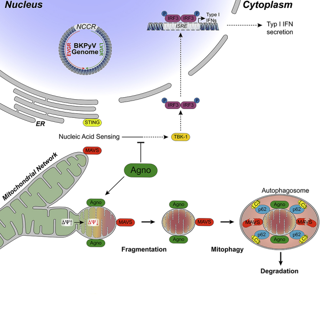
          Immune escape contributes to viral persistence, yet little is known about human polyomaviruses. BK-polyomavirus (BKPyV) asymptomatically infects 90% of humans but causes premature allograft failure in kidney transplant patients. Despite virus-specific T cells and neutralizing antibodies, BKPyV persists in kidneys and evades immune control as evidenced by urinary shedding in immunocompetent individuals. Here, we report that BKPyV disrupts the mitochondrial network and membrane potential when expressing the 66aa-long agnoprotein during late replication. Agnoprotein is necessary and sufficient, using its amino-terminal and central domain for mitochondrial targeting and network disruption, respectively. Agnoprotein impairs nuclear IRF3-translocation, interferon-beta expression, and promotes p62/SQSTM1-mitophagy. Agnoprotein-mutant viruses unable to disrupt mitochondria show reduced replication and increased interferon-beta expression but can be rescued by type-I interferon blockade, TBK1-inhibition, or CoCl 2-treatment. Mitochondrial fragmentation and p62/SQSTM1-autophagy occur in allograft biopsies of kidney transplant patients with BKPyV nephropathy. JCPyV and SV40 infection similarly disrupt mitochondrial networks, indicating a conserved mechanism facilitating polyomavirus persistence and post-transplant disease.

          Graphical Abstract

          Highlights

          • BK polyomavirus agnoprotein disrupts mitochondrial membrane potential and network

          • Agnoprotein impairs nucleus IRF3 translocation and interferon-β expression

          • Agnoprotein facilitates innate immune evasion during the late viral replication phase

          • Damaged mitochondria are targeted for p62/SQSTM1 autophagy

          Abstract

          Biological Sciences; Immunology; Virology; Cell Biology

          Related collections

          Most cited references63

          • Record: found
          • Abstract: found
          • Article: not found

          DNA damage primes the type I interferon system via the cytosolic DNA sensor STING to promote anti-microbial innate immunity.

          Dysfunction in Ataxia-telangiectasia mutated (ATM), a central component of the DNA repair machinery, results in Ataxia Telangiectasia (AT), a cancer-prone disease with a variety of inflammatory manifestations. By analyzing AT patient samples and Atm(-/-) mice, we found that unrepaired DNA lesions induce type I interferons (IFNs), resulting in enhanced anti-viral and anti-bacterial responses in Atm(-/-) mice. Priming of the type I interferon system by DNA damage involved release of DNA into the cytoplasm where it activated the cytosolic DNA sensing STING-mediated pathway, which in turn enhanced responses to innate stimuli by activating the expression of Toll-like receptors, RIG-I-like receptors, cytoplasmic DNA sensors, and their downstream signaling partners. This study provides a potential explanation for the inflammatory phenotype of AT patients and establishes damaged DNA as a cell intrinsic danger signal that primes the innate immune system for a rapid and amplified response to microbial and environmental threats.
            Bookmark
            • Record: found
            • Abstract: found
            • Article: not found

            Selective autophagy mediated by autophagic adapter proteins.

            Mounting evidence suggests that autophagy is a more selective process than originally anticipated. The discovery and characterization of autophagic adapters, like p62 and NBR1, has provided mechanistic insight into this process. p62 and NBR1 are both selectively degraded by autophagy and able to act as cargo receptors for degradation of ubiquitinated substrates. A direct interaction between these autophagic adapters and the autophagosomal marker protein LC3, mediated by a so-called LIR (LC3-interacting region) motif, their inherent ability to polymerize or aggregate as well as their ability to specifically recognize substrates are required for efficient selective autophagy. These three required features of autophagic cargo receptors are evolutionarily conserved and also employed in the yeast cytoplasm-to-vacuole targeting (Cvt) pathway and in the degradation of P granules in C. elegans. Here, we review the mechanistic basis of selective autophagy in mammalian cells discussing the degradation of misfolded proteins, p62 bodies, aggresomes, mitochondria and invading bacteria. The emerging picture of selective autophagy affecting the regulation of cell signaling with consequences for oxidative stress responses, tumorigenesis and innate immunity is also addressed.
              Bookmark
              • Record: found
              • Abstract: found
              • Article: not found

              The virome in mammalian physiology and disease.

              The virome contains the most abundant and fastest mutating genetic elements on Earth. The mammalian virome is constituted of viruses that infect host cells, virus-derived elements in our chromosomes, and viruses that infect the broad array of other types of organisms that inhabit us. Virome interactions with the host cannot be encompassed by a monotheistic view of viruses as pathogens. Instead, the genetic and transcriptional identity of mammals is defined in part by our coevolved virome, a concept with profound implications for understanding health and disease. Copyright © 2014 Elsevier Inc. All rights reserved.
                Bookmark

                Author and article information

                Contributors
                Journal
                iScience
                iScience
                iScience
                Elsevier
                2589-0042
                10 June 2020
                24 July 2020
                10 June 2020
                : 23
                : 7
                : 101257
                Affiliations
                [1 ]Transplantation & Clinical Virology, Department Biomedicine (Haus Petersplatz), University of Basel, Petersplatz 10, CH-4009 Basel, Switzerland
                [2 ]Institute for Pathology, University Hospital Basel, Basel, Switzerland
                [3 ]Departments of Pathology, University of Maryland School of Medicine, Baltimore, MD, USA
                [4 ]Department of Microbiology and Infection Control, University Hospital of North Norway, Tromsø, Norway
                [5 ]Metabolic and Renal Research Group, UiT The Arctic University of Norway, Tromsø, Norway
                [6 ]Clinical Virology, Laboratory Medicine, University Hospital Basel, Basel, Switzerland
                [7 ]Infectious Diseases & Hospital Epidemiology, University Hospital Basel, Basel, Switzerland
                Author notes
                []Corresponding author hans.hirsch@ 123456unibas.ch
                [8]

                Present address: Cancer Immunotherapy, Department Biomedicine, University of Basel, Basel, Switzerland

                [9]

                Present address: Immunobiology, Department Biomedicine, University of Basel, Basel, Switzerland

                [10]

                Lead contact

                Article
                S2589-0042(20)30443-0 101257
                10.1016/j.isci.2020.101257
                7326741
                32599557
                7b0b8dc5-469d-46a6-89d0-5ee30fcd6938
                © 2020 The Author(s)

                This is an open access article under the CC BY-NC-ND license (http://creativecommons.org/licenses/by-nc-nd/4.0/).

                History
                : 10 September 2019
                : 16 March 2020
                : 5 June 2020
                Categories
                Article

                biological sciences,immunology,virology,cell biology
                biological sciences, immunology, virology, cell biology

                Comments

                Comment on this article