0
views
0
recommends
+1 Recommend
0 collections
    0
    shares
      • Record: found
      • Abstract: found
      • Article: found
      Is Open Access

      dsRNA-induced condensation of antiviral proteins modulates PKR activity

      research-article

      Read this article at

      Bookmark
          There is no author summary for this article yet. Authors can add summaries to their articles on ScienceOpen to make them more accessible to a non-specialist audience.

          Significance

          The presence of dsRNA in the cytosol is a marker of infection and elicits an immune response. One aspect of this immune response is the activation of the eIF2α kinase PKR, which results in translational reprogramming and stress granule formation. Here, we show that dsRNA induces the formation of a novel condensate by PKR that is distinct from other known ribonucleoprotein assemblies. These results challenge prior observations that PKR is recruited to stress granules and suggest that the condensation of PKR may be a mechanism that cells use to modulate PKR activation.

          Abstract

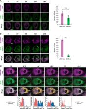
          Mammalian cells respond to dsRNA in multiple manners. One key response to dsRNA is the activation of PKR, an eIF2α kinase, which triggers translational arrest and the formation of stress granules. However, the process of PKR activation in cells is not fully understood. In response to increased endogenous or exogenous dsRNA, we observed that PKR forms novel cytosolic condensates, referred to as dsRNA-induced foci (dRIFs). dRIFs contain dsRNA, form in proportion to dsRNA, and are enhanced by longer dsRNAs. dRIFs enrich several other dsRNA-binding proteins, including ADAR1, Stau1, NLRP1, and PACT. Strikingly, dRIFs correlate with and form before translation repression by PKR and localize to regions of cells where PKR activation is initiated. We hypothesize that dRIF formation is a mechanism that cells use to enhance the sensitivity of PKR activation in response to low levels of dsRNA or to overcome viral inhibitors of PKR activation.

          Related collections

          Most cited references58

          • Record: found
          • Abstract: found
          • Article: not found

          Pattern recognition receptors and inflammation.

          Infection of cells by microorganisms activates the inflammatory response. The initial sensing of infection is mediated by innate pattern recognition receptors (PRRs), which include Toll-like receptors, RIG-I-like receptors, NOD-like receptors, and C-type lectin receptors. The intracellular signaling cascades triggered by these PRRs lead to transcriptional expression of inflammatory mediators that coordinate the elimination of pathogens and infected cells. However, aberrant activation of this system leads to immunodeficiency, septic shock, or induction of autoimmunity. In this Review, we discuss the role of PRRs, their signaling pathways, and how they control inflammatory responses. 2010 Elsevier Inc. All rights reserved.
            Bookmark
            • Record: found
            • Abstract: found
            • Article: not found

            ATPase-Modulated Stress Granules Contain a Diverse Proteome and Substructure.

            Stress granules are mRNA-protein granules that form when translation initiation is limited, and they are related to pathological granules in various neurodegenerative diseases. Super-resolution microscopy reveals stable substructures, referred to as cores, within stress granules that can be purified. Proteomic analysis of stress granule cores reveals a dense network of protein-protein interactions and links between stress granules and human diseases and identifies ATP-dependent helicases and protein remodelers as conserved stress granule components. ATP is required for stress granule assembly and dynamics. Moreover, multiple ATP-driven machines affect stress granules differently, with the CCT complex inhibiting stress granule assembly, while the MCM and RVB complexes promote stress granule persistence. Our observations suggest that stress granules contain a stable core structure surrounded by a dynamic shell with assembly, disassembly, and transitions between the core and shell modulated by numerous protein and RNA remodeling complexes.
              Bookmark
              • Record: found
              • Abstract: found
              • Article: not found

              Compositional Control of Phase-Separated Cellular Bodies.

              Cellular bodies such as P bodies and PML nuclear bodies (PML NBs) appear to be phase-separated liquids organized by multivalent interactions among proteins and RNA molecules. Although many components of various cellular bodies are known, general principles that define body composition are lacking. We modeled cellular bodies using several engineered multivalent proteins and RNA. In vitro and in cells, these scaffold molecules form phase-separated liquids that concentrate low valency client proteins. Clients partition differently depending on the ratio of scaffolds, with a sharp switch across the phase diagram diagonal. Composition can switch rapidly through changes in scaffold concentration or valency. Natural PML NBs and P bodies show analogous partitioning behavior, suggesting how their compositions could be controlled by levels of PML SUMOylation or cellular mRNA concentration, respectively. The data suggest a conceptual framework for considering the composition and control thereof of cellular bodies assembled through heterotypic multivalent interactions.
                Bookmark

                Author and article information

                Journal
                Proc Natl Acad Sci U S A
                Proc Natl Acad Sci U S A
                pnas
                PNAS
                Proceedings of the National Academy of Sciences of the United States of America
                National Academy of Sciences
                0027-8424
                1091-6490
                8 August 2022
                16 August 2022
                8 August 2022
                : 119
                : 33
                : e2204235119
                Affiliations
                [1] aDepartment of Biochemistry, University of Colorado , Boulder, CO 80309;
                [2] bBioFrontiers Institute , Boulder, CO 80309;
                [3] cHoward Hughes Medical Institute , Chevy Chase, MD 20815-6789
                Author notes
                1To whom correspondence may be addressed. Email: roy.parker@ 123456colorado.edu .

                Contributed by Roy Parker; received March 10, 2022; accepted July 1, 2022; reviewed by Diego Acosta-Alvear and Hao Wu

                Author contributions: G.A.C., J.M.B., and R.P. designed research; G.A.C. and G.R.B. performed research; G.A.C. and J.W.T. analyzed data; and G.A.C., J.M.B., J.W.T., and R.P. wrote the paper.

                Author information
                https://orcid.org/0000-0002-8412-4152
                Article
                202204235
                10.1073/pnas.2204235119
                9388085
                35939694
                79dab491-9d22-4fb8-87fc-2cf01ebf1964
                Copyright © 2022 the Author(s). Published by PNAS.

                This open access article is distributed under Creative Commons Attribution License 4.0 (CC BY).

                History
                : 01 July 2022
                Page count
                Pages: 12
                Funding
                Funded by: HHS | National Institutes of Health (NIH) 100000002
                Award ID: GM045443
                Award Recipient : Giulia A Corbet Award Recipient : James M Burke Award Recipient : Gaia R Bublitz Award Recipient : Roy Parker
                Funded by: Howard Hughes Medical Institute (HHMI) 100000011
                Award ID: N/A
                Award Recipient : Roy Parker
                Categories
                409
                Biological Sciences
                Cell Biology

                dsrna,pkr,condensate
                dsrna, pkr, condensate

                Comments

                Comment on this article