0
views
0
recommends
+1 Recommend
0 collections
    0
    shares
      • Record: found
      • Abstract: found
      • Article: found
      Is Open Access

      Ang Ⅱ诱导大鼠心房肌纤维化的mRNA表达谱变化 Translated title: mRNA Expression Profile Changes in Angiotensin-Ⅱ-Induced Atrial Myocardial Fibrosis in Rats

      research-article

      Read this article at

      Bookmark
          There is no author summary for this article yet. Authors can add summaries to their articles on ScienceOpen to make them more accessible to a non-specialist audience.

          Abstract

          目的

          研究血管紧张素Ⅱ(Ang Ⅱ)诱导的大鼠纤维化心肌细胞与正常大鼠心肌细胞mRNA表达谱之间的差异及其相关信号通路。

          方法

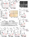
          8周龄雄性SD大鼠6只,随机分为对照组(Control组)和Ang Ⅱ组,每组3只。Control组每日经尾静脉注射生理盐水,Ang Ⅱ组则注射2 mg/kg的Ang Ⅱ,两组持续给药14 d。采用Masson染色法检测大鼠心肌纤维化程度,免疫组化法检测胶原蛋白Ⅰ含量。利用高通量测序技术检测两组大鼠心肌细胞mRNA的表达并筛选出差异mRNA,进行GO分析和KEGG通路分析。

          结果

          与Control组相比,Ang Ⅱ组的心肌纤维化程度及胶原蛋白Ⅰ含量升高( P<0.05)。进行测序后共筛选出313条差异mRNA(其中201条上调,112条下调)。GO和KEGG分析表明上述差异表达的mRNA参与了心肌纤维化的多种生物学调节功能和通路。

          结论

          Ang Ⅱ可以导致大鼠心肌发生纤维化;纤维化的心肌细胞与正常心肌细胞之间的mRNA表达具有明显差异,差异表达的mRNA在免疫反应、细胞重构、细胞外基质沉积等生物过程中可能发挥重要作用。

          Translated abstract

          Objective

          To study the differences between the mRNA expression profile in angiotensin Ⅱ (Ang Ⅱ)-induced fibrotic cardiomyocytes and that of normal cardiomyocytes and the relevant signaling pathways.

          Methods

          Six 8-week-old male Sprague-Dawley (SD) rats were randomly assigned to a control group and an Ang Ⅱ group, with 3 rats in each group. Rats in the control group were injected via caudal vein with 0.9% normal saline at 2 mg/kg per day, while rats in the Ang Ⅱ group were injected with Ang Ⅱ via caudal vein at 2 mg/kg per day. The medications were continuously administered in the two groups for 14 days. The degree of myocardial fibrosis was determined by Masson's Trichrome staining and the content of collagen Ⅰ was determined by immunohistochemistry. High throughput sequencing was performed to measure the mRNA expression of rat cardiomyocytes in the two groups and to screen for differentially-expressed mRNAs. The differentially-expressed mRNAs were analyzed by Gene Ontology (GO) analysis and Kyoto Encyclopedia of Genes and Genomes (KEGG) pathway analysis.

          Results

          Compared with those of the control group, the degree of myocardial fibrosis and the content of collagen Ⅰ in Ang Ⅱ group were significantly higher ( P<0.05). Through sequencing, 313 differentially-expressed mRNAs were identified, with 201 being up-regulated and 112 being down-regulated. Go and KEGG analyses showed that these differentially-expressed mRNA were involved in a variety of biological regulatory functions and pathways of myocardial fibrosis.

          Conclusion

          Ang Ⅱ can cause myocardial fibrosis in rats. There are significant differences in mRNA expression between fibrotic cardiomyocytes and normal cardiomyocytes. The differentially expressed mRNAs may play an important role in biological processes, including immune response, cell remodeling, and extracellular matrix deposition.

          Related collections

          Most cited references20

          • Record: found
          • Abstract: found
          • Article: not found

          Atrial fibrillation and cardiac fibrosis

          The understanding of atrial fibrillation (AF) evolved from a sole rhythm disturbance towards the complex concept of a cardiomyopathy based on arrhythmia substrates. There is evidence that atrial fibrosis can be visualized using late gadolinium enhancement cardiac magnetic resonance imaging and that it is a powerful predictor for the outcome of AF interventions. However, a strategy of an individual and fibrosis guided management of AF looks promising but results from prospective multicentre trials are pending. This review gives an overview about the relationship between cardiac fibrosis and AF focusing on translational aspects, clinical observations, and fibrosis imaging to emphasize the concept of personalized paths in AF management taking into account the individual amount and distribution of fibrosis.
            Bookmark
            • Record: found
            • Abstract: found
            • Article: found
            Is Open Access

            Blockade of Fibroblast YAP Attenuates Cardiac Fibrosis and Dysfunction Through MRTF-A Inhibition

            Visual Abstract
              Bookmark
              • Record: found
              • Abstract: found
              • Article: found
              Is Open Access

              Overexpression of miR-27b-3p Targeting Wnt3a Regulates the Signaling Pathway of Wnt/ β -Catenin and Attenuates Atrial Fibrosis in Rats with Atrial Fibrillation

              MicroRNAs (miRNAs) are regarded as a potential method for the treatment of atrial fibrillation (AF) although its molecular mechanism remains unknown. We found in our previous study that the level of peripheral blood miR-27b-3p and the expression of atrial tissue CX43 were both significantly downregulated in AF patients. In the present study, we propose and test this hypothesis that overexpression of miR-27b-3p attenuates atrial fibrosis, increases CX43 expression, and regulates the signaling pathway of Wnt/β-Catenin by targeting Wnt3a. miR-27b-3p overexpression was induced by rat tail vein injection of adeno-associated virus. Two weeks after transfection of adeno-associated virus, the rat AF model was established by tail vein injection of acetylcholine- (ACh-) CaCl2 for 7 days, and 1 ml/kg was injected daily. The incidence and duration of AF were recorded with an electrocardiogram. Cardiac function was monitored by cardiac ultrasound. Serum cardiac enzyme was detected by ELISA. The expression of atrial miR-27b-3 and Wnt3a was assayed by quantitative RT-PCR. Atrial fibrosis was determined by Masson's trichrome staining. Expression of atrial Collagen-I and Collagen-III was tested by the immunohistochemical method. Expression of CX43 was measured by immunofluorescence. The expression of Collagen-I, a-SMA, Collagen-III, TGF-β1, CX43, Wnt3a, β-Catenin, and p-β-Catenin was assayed by western blot. Our results showed that miR-27b-3p overexpression could reduce the incidence and duration of AF, alleviate atrial fibrosis, increase atrial CX43 expression, and decrease the expression of Collagen-I, a-SMA, Collagen-III, TGF-β1, Wnt3a, and p-β-Catenin. In addition, the results of luciferase activity assay showed that Wnt3a is a validated miR-27b-3p target in HEK 293T cells. Our results provide a new evidence that miR-27b-3p regulates the signaling pathway of Wnt/β-Catenin by targeting Wnt3a, which may play an important role in the development of atrial fibrosis and AF.
                Bookmark

                Author and article information

                Contributors
                Journal
                Sichuan Da Xue Xue Bao Yi Xue Ban
                Sichuan Da Xue Xue Bao Yi Xue Ban
                SCDXXBYXB
                Journal of Sichuan University (Medical Sciences)
                四川大学学报(医学版)编辑部 (中国四川 )
                1672-173X
                20 September 2023
                : 54
                : 5
                : 959-964
                Affiliations
                [1 ] 桂林医学院附属医院 综合科医疗保健病区 (桂林 541001) Department of Medical Care Ward, Affiliated Hospital of Guilin Medical University, Guilin 541001, China
                [2 ] 桂林医学院 发展规划和医院管理处 (桂林 541199) Department of Development Planning and Hospital Management, Guilin Medical University, Guilin 541199, China
                [3 ] 桂林医学院 (桂林 541199) Guilin Medical University, Guilin 541199, China
                [4 ] 桂林医学院附属医院 组织科 (桂林 541001) Department of Organization, Affiliated Hospital of Guilin Medical University, Guilin 541001, China
                Author notes
                Article
                scdxxbyxb-54-5-959
                10.12182/20230960211
                10579065
                79399ed1-dd98-4446-aeec-9924cef42d9c
                © 2023《四川大学学报(医学版)》编辑部 版权所有Copyright ©2023 Editorial Board of Journal of Sichuan University (Medical Sciences)

                开放获取 本文遵循知识共享署名—非商业性使用4.0国际许可协议(CC BY-NC 4.0),允许第三方对本刊发表的论文自由共享(即在任何媒介以任何形式复制、发行原文)、演绎(即修改、转换或以原文为基础进行创作),必须给出适当的署名,提供指向本文许可协议的链接,同时标明是否对原文作了修改;不得将本文用于商业目的。CC BY-NC 4.0许可协议访问 https://creativecommons.org/licenses/by-nc/4.0

                Open Access This article is licensed under a Creative Commons Attribution-NonCommercial 4.0 International license (CC BY-NC 4.0). In other words, the full-text content of the journal is made freely available for third-party users to copy and redistribute in any medium or format, and to remix, transform, and build upon the content of the journal. You must give appropriate credit, provide a link to the license, and indicate if changes were made. You may not use the content of the journal for commercial purposes. For more information about the license, visit https://creativecommons.org/licenses/by-nc/4.0

                History
                : 3 February 2023
                Funding
                国家自然科学基金地区项目(No. 82160068)和国家级大学生创新创业训练计划项目资助(No. 202110601004)资助
                Categories
                论 著

                血管紧张素ⅱ,心肌纤维化,mrna,angiotensin ⅱ,myocardioal fibrosis

                Comments

                Comment on this article