87
views
0
recommends
+1 Recommend
0 collections
    0
    shares
      • Record: found
      • Abstract: found
      • Article: found
      Is Open Access

      Cell-of-Origin Patterns Dominate the Molecular Classification of 10,000 Tumors from 33 Types of Cancer

      research-article

      Read this article at

      Bookmark
          There is no author summary for this article yet. Authors can add summaries to their articles on ScienceOpen to make them more accessible to a non-specialist audience.

          SUMMARY

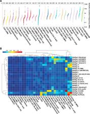
          We conducted comprehensive integrative molecular analyses of the complete set of tumors in The Cancer Genome Atlas (TCGA), consisting of approximately 10,000 specimens and representing 33 types of cancer. We performed molecular clustering using data on chromosome-arm-level aneuploidy, DNA hypermethylation, mRNA, and miRNA expression levels and reverse-phase protein arrays, of which all, except for aneuploidy, revealed clustering primarily organized by histology, tissue type, or anatomic origin. The influence of cell type was evident in DNA-methylation-based clustering, even after excluding sites with known preexisting tissue-type-specific methylation. Integrative clustering further emphasized the dominant role of cell-of-origin patterns. Molecular similarities among histologically or anatomically related cancer types provide a basis for focused pan-cancer analyses, such as pan-gastrointestinal, pan-gynecological, pan-kidney, and pan-squamous cancers, and those related by stemness features, which in turn may inform strategies for future therapeutic development.

          In Brief

          Comprehensive, integrated molecular analysis identifies molecular relationships across a large diverse set of human cancers, suggesting future directions for exploring clinical actionability in cancer treatment.

          Related collections

          Most cited references20

          • Record: found
          • Abstract: found
          • Article: found

          Comprehensive and Integrative Genomic Characterization of Hepatocellular Carcinoma

          (2017)
          Liver cancer has the second highest worldwide cancer mortality rate and has limited therapeutic options. We analyzed 363 hepatocellular carcinoma (HCC) cases by whole exome sequencing and DNA copy number analyses, and 196 HCC also by DNA methylation, RNA, miRNA, and proteomic expression. DNA sequencing and mutation analysis identified significantly mutated genes including LZTR1 , EEF1A1 , SF3B1 , and SMARCA4 . Significant alterations by mutation or down-regulation by hypermethylation in genes likely to result in HCC metabolic reprogramming ( ALB , APOB , and CPS1 ) were observed. Integrative molecular HCC subtyping incorporating unsupervised clustering of five data platforms identified three subtypes, one of which was associated with poorer prognosis in three HCC cohorts. Integrated analyses enabled development of a p53 target gene expression signature correlating with poor survival. Potential therapeutic targets for which inhibitors exist include WNT signaling, MDM4, MET, VEGFA, MCL1, IDH1, TERT, and immune checkpoint proteins CTLA-4, PD-1, and PD-L1. Multiplex molecular profiling of human hepatocellular carcinoma patients provides insight into subtype characteristics and points toward key pathways to target therapeutically.
            Bookmark
            • Record: found
            • Abstract: found
            • Article: not found

            Multiplatform analysis of 12 cancer types reveals molecular classification within and across tissues of origin.

            Recent genomic analyses of pathologically defined tumor types identify "within-a-tissue" disease subtypes. However, the extent to which genomic signatures are shared across tissues is still unclear. We performed an integrative analysis using five genome-wide platforms and one proteomic platform on 3,527 specimens from 12 cancer types, revealing a unified classification into 11 major subtypes. Five subtypes were nearly identical to their tissue-of-origin counterparts, but several distinct cancer types were found to converge into common subtypes. Lung squamous, head and neck, and a subset of bladder cancers coalesced into one subtype typified by TP53 alterations, TP63 amplifications, and high expression of immune and proliferation pathway genes. Of note, bladder cancers split into three pan-cancer subtypes. The multiplatform classification, while correlated with tissue-of-origin, provides independent information for predicting clinical outcomes. All data sets are available for data-mining from a unified resource to support further biological discoveries and insights into novel therapeutic strategies. Copyright © 2014 Elsevier Inc. All rights reserved.
              Bookmark
              • Record: found
              • Abstract: found
              • Article: not found

              JAK–STAT Signaling as a Target for Inflammatory and Autoimmune Diseases: Current and Future Prospects

              The Janus kinase/signal transduction and activator of transcription (JAK–STAT) signaling pathway is implicated in the pathogenesis of inflammatory and autoimmune diseases including rheumatoid arthritis, psoriasis, and inflammatory bowel disease. Many cytokines involved in the pathogenesis of autoimmune and inflammatory diseases use JAKs and STATs to transduce intracellular signals. Mutations in JAK and STAT genes cause a number of immunodeficiency syndromes, and polymorphisms in these genes are associated with autoimmune diseases. The success of small-molecule JAK inhibitors (Jakinibs) in the treatment of rheumatologic disease demonstrates that intracellular signaling pathways can be targeted therapeutically to treat autoimmunity. Tofacitinib, the first rheumatologic Jakinib, is US Food and Drug Administration (FDA) approved for rheumatoid arthritis and is currently under investigation for other autoimmune diseases. Many other Jakinibs are in preclinical development or in various phases of clinical trials. This review describes the JAK–STAT pathway, outlines its role in autoimmunity, and explains the rationale/pre-clinical evidence for targeting JAK–STAT signaling. The safety and clinical efficacy of the Jakinibs are reviewed, starting with the FDA-approved Jakinib tofacitinib, and continuing on to next-generation Jakinibs. Recent and ongoing studies are emphasized, with a focus on emerging indications for JAK inhibition and novel mechanisms of JAK–STAT signaling blockade.
                Bookmark

                Author and article information

                Journal
                0413066
                2830
                Cell
                Cell
                Cell
                0092-8674
                1097-4172
                17 April 2018
                05 April 2018
                05 April 2019
                : 173
                : 2
                : 291-304.e6
                Affiliations
                [1 ]Department of Genetics, Lineberger Comprehensive Cancer Center, the University of North Carolina at Chapel Hill, Chapel Hill, NC 27599, USA
                [2 ]Buck Institute for Research on Aging, Novato, CA 94945, USA
                [3 ]Department of Surgery, University of California, San Francisco, San Francisco, CA 94115, USA
                [4 ]Van Andel Research Institute, Grand Rapids, MI 49503, USA
                [5 ]Department of Laboratory Medicine, University of California, San Francisco, San Francisco, CA 94115, USA
                [6 ]Department of Bioinformatics and Computational Biology, The University of Texas MD Anderson Cancer Center, Houston, TX 77030, USA
                [7 ]Department of Epidemiology and Biostatistics, Memorial Sloan Kettering Cancer Center, New York, NY 10065, USA
                [8 ]Broad Institute of Harvard and MIT, Cambridge, MA 02142, USA
                [9 ]Department of Medical Oncology, Dana-Farber Cancer Institute, Boston, MA 02215, USA
                [10 ]Institute for Systems Biology, Seattle, WA 98109, USA
                [11 ]Canada’s Michael Smith Genome Sciences Centre, BC Cancer Agency, Vancouver, BC V5Z 1L3, Canada
                [12 ]Department of Biomolecular Engineering, Center for Biomolecular Sciences and Engineering, University of California, Santa Cruz, Santa Cruz, CA 95064, USA
                [13 ]Poznań University of Medical Sciences, 61-701 Poznań, Poland
                [14 ]Greater Poland Cancer Centre, 61-866 Poznań, Poland
                [15 ]International Institute for Molecular Oncology, 60-203 Poznań, Poland
                [16 ]Marie-Josée and Henry R. Kravis Center for Molecular Oncology, Memorial Sloan Kettering Cancer Center, New York, NY 10065, USA
                [17 ]Department of Medicine, Division of Gastroenterology, Vanderbilt University Medical Center, Nashville, TN 37232, USA
                [18 ]Massachusetts General Hospital Cancer Center and Department of Pathology, Harvard Medical School, Charlestown, MA 02129, USA
                [19 ]Department of Neurosurgery, Henry Ford Health System, Detroit, MI 48202, USA
                [20 ]Department of Genetics, University of Sao Paulo, Ribeirao Preto, SP, 14049-900, Brazil
                Author notes
                [* ]Correspondence: hoadley@ 123456med.unc.edu (K.A.H.), peter.laird@ 123456vai.org (P.W.L.)
                [21]

                These authors contributed equally

                [22]

                Lead Contact

                Article
                NIHMS957746
                10.1016/j.cell.2018.03.022
                5957518
                29625048
                7751268d-5047-4d7a-9e25-93681842b6a2

                This is an open access article under the CC BY-NC-ND license ( http://creativecommons.org/licenses/by-nc-nd/4.0/).

                History
                Categories
                Article

                Cell biology
                Cell biology

                Comments

                Comment on this article