10
views
0
recommends
+1 Recommend
0 collections
    0
    shares
      • Record: found
      • Abstract: found
      • Article: found
      Is Open Access

      MULTIZONAL OUTER RETINOPATHY AND RETINAL PIGMENT EPITHELIOPATHY (MORR) : A Newly Recognized Entity or an Unusual Variant of AZOOR?

      research-article

      Read this article at

      Bookmark
          There is no author summary for this article yet. Authors can add summaries to their articles on ScienceOpen to make them more accessible to a non-specialist audience.

          Abstract

          This longitudinal description of acute zonal occult outer retinopathy patients over a long-term period using multimodal imaging uncovered an unusual entity characterized by multizonal involvement and a prolonged progressive clinical course.

          Abstract

          Purpose:

          To describe specific clinical, multimodal imaging, and natural history features of an unusual variant of acute zonal occult outer retinopathy.

          Methods:

          Retrospective, observational, longitudinal, multicenter case series. Patients exhibiting this unusual clinical condition among cases previously diagnosed with acute zonal occult outer retinopathy were included. Multimodal imaging, laboratory evaluations, and genetic testing for inherited retinal diseases were reviewed.

          Results:

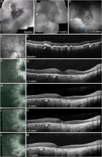
          Twenty eyes from 10 patients (8 females and 2 males) with a mean age of 54.1 ± 13.3 years (range, 38–71 years) were included. The mean follow-up duration was 13.1 ± 5.3 years (range, 8–23 years). Presenting symptoms were bilateral in 7 patients (85% of eyes) and included scotomata and photopsia. All patients had bilateral lesions at presentation involving the peripapillary and far peripheral retina. Baseline optical coherence tomography showed alteration of the retinal pigment epithelium and photoreceptor layers corresponding to zonal areas of fundus autofluorescence abnormalities. Centrifugal and centripetal progression of the peripapillary and far-peripheral lesions, respectively, occurred over the follow-up, resulting in areas of complete outer retinal and retinal pigment epithelium atrophy.

          Conclusion:

          Initial alteration of photoreceptors and retinal pigment epithelium and a stereotypical natural course that includes involvement of the far retinal periphery, characterize this unusual condition. It may represent a variant of acute zonal occult outer retinopathy or may be a new entity. We suggest to call it multizonal outer retinopathy and retinal pigment epitheliopathy.

          Related collections

          Most cited references23

          • Record: found
          • Abstract: found
          • Article: not found

          Acute zonal occult outer retinopathy: a long-term follow-up study.

          To report the long-term follow-up of patients with acute zonal occult outer retinopathy (AZOOR). Observational consecutive case series. Prospective and retrospective review of medical records of patients with AZOOR. Fifty-one patients (37 women and 14 men) with a median age of 33 years (mean, age 36 years; range, 13-63 years) were followed for a median of 96 months (mean 100 months; range, 36-420 months). At presentation, AZOOR was present in one eye of 31 patients (61%) and both eyes in 20 patients (39%). All patients presented with an acute loss of one or more zones of visual field, and 45 (88%) patients presented with photopsia. Corrected visual acuity was 20/40 or better in 68 (76%) of 90 affected eyes. Funduscopic examination was normal in 82 eyes and revealed signs of AZOOR in 8 eyes. Electroretinographic amplitudes were depressed in all affected eyes. The median delay in diagnosis of AZOOR was 17 months. During follow-up, AZOOR developed in 19 fellow eyes. At final follow-up AZOOR was present in one eye of 12 (24%) patients and both eyes of 39 (76%) patients. Sixteen patients had 23 recurrences of AZOOR. Visual field loss stabilized within 6 months in 37 patients (72%), progressed stepwise in 2 patients (4%), and partly improved in 12 patients (24%). Fourteen patients (28%) had autoimmune diseases, including Hashimoto's thyroiditis in 6 patients and relapsing transverse myelopathy in 4 patients. At last follow-up all patients had residual visual field defects. Final visual acuity was 20/40 or better in 61 (68%) affected eyes. Nine patients (18%) were legally blind. The fundi of 90 affected eyes revealed no changes of AZOOR in 47 eyes (52%) and changes in the pigment epithelium and retina caused by AZOOR in 43 eyes (48%). Visual loss in AZOOR is characterized by one or more episodes of acute dysfunction, and in some cases, death of retinal receptor cells in one or more zones of one or both eyes. Central vision is often spared, but recovery of visual field occurs infrequently. The etiology of AZOOR is unknown. Electroretinography is essential for early diagnosis. The value of treatment is uncertain.
            Bookmark
            • Record: found
            • Abstract: not found
            • Article: not found

            White spot syndromes of the retina: a hypothesis based on the common genetic hypothesis of autoimmune/inflammatory disease.

              Bookmark
              • Record: found
              • Abstract: found
              • Article: not found

              Acute zonal occult outer retinopathy. Donders Lecture: The Netherlands Ophthalmological Society, Maastricht, Holland, June 19, 1992.

              J Gass (1993)
              This report describes 13 patients, predominantly young women, with a syndrome characterized by rapid loss of one or more large zones of outer retinal function, photopsia, minimal funduscopic changes, and electroretinographic abnormalities affecting one or both eyes. All patients on follow-up examination had persistent visual field defects, and most had chronic photopsia and zones of pigment epithelial atrophy. Evidence is presented that these patients probably represent part of the spectrum of a single disorder that includes the multiple evanescent white-dot syndrome (MEWDS), acute idiopathic blind-spot-enlargement syndrome (AIBSES), acute macular neuroretinopathy (AMN), and the pseudo-presumed ocular histoplasmosis syndrome (P-POHS). The medical records of these 13 patients and 2 additional patients, who developed, in addition to the features of this syndrome, funduscopic changes typical of MEWDS, AMN, and P-POHS, were reviewed and follow-up obtained. These patients had extensive unrewarding medical and neurological investigations because of suspected diagnoses, including central nervous system disorders, cancer-associated retinopathy, retinal vasculitis, diffuse unilateral subacute neuroretinitis, and tapetoretinal degenerations. Although most patients retained good visual acuity, all had permanent visual field loss that in some cases was severe. The cause of the disorder was not determined. No effective treatment was found. Acute visual loss and photopsia in these patients is probably caused by damage to large zones of the outer retina that appears unaffected ophthalmoscopically. Electroretinography is important in early diagnosis. Future investigations probably will reveal further evidence linking this disorder to MEWDS, AIBSES, AMN, and P-POHS.
                Bookmark

                Author and article information

                Contributors
                Journal
                Retina
                Retina
                retina
                Retina (Philadelphia, Pa.)
                Retina
                0275-004X
                1539-2864
                November 2023
                19 October 2023
                : 43
                : 11
                : 1890-1903
                Affiliations
                [* ]Vitreous Retina Macula Consultants of New York, New York, NY;
                []Department of Ophthalmology, Feinberg School of Medicine, Northwestern University, Chicago, IL;
                []Department of Ophthalmology, IRCCS San Raffaele Scientific Institute, Milan, Italy;
                [§ ]School of Medicine, Vita-Salute San Raffaele University, Milan, Italy; and
                []Department of Ophthalmology, NYU Grossman School of Medicine, New York, NY.
                Author notes
                Reprint requests: Prithvi Ramtohul, MD, Vitreous Retina Macula Consultants of New York, 950 Third Avenue, New York, NY 10022; e-mail: pramtohul@ 123456me.com
                Article
                RETINA-223-0644 00008
                10.1097/IAE.0000000000003927
                10589432
                37748093
                7712d7b7-3acb-430d-9cea-8eb040f65ffa
                Copyright © 2023 The Author(s). Published by Wolters Kluwer Health, Inc. on behalf of the Opthalmic Communications Society, Inc.

                This is an open access article distributed under the terms of the Creative Commons Attribution-Non Commercial-No Derivatives License 4.0 (CCBY-NC-ND), where it is permissible to download and share the work provided it is properly cited. The work cannot be changed in any way or used commercially without permission from the journal.

                History
                Categories
                Original Study
                Custom metadata
                TRUE
                T
                podcast

                azoor,multimodal imaging,optical coherence tomography,morr,multizonal outer retinopathy and retinal pigment epitheliopathy

                Comments

                Comment on this article