53
views
0
recommends
+1 Recommend
0 collections
    0
    shares
      • Record: found
      • Abstract: found
      • Article: not found

      NAG-1/GDF15 prevents obesity by increasing thermogenesis, lipolysis and oxidative metabolism

      research-article

      Read this article at

      ScienceOpenPublisherPMC
      Bookmark
          There is no author summary for this article yet. Authors can add summaries to their articles on ScienceOpen to make them more accessible to a non-specialist audience.

          Summary

          Objective

          Obesity is a major health problem associated with high morbidity and mortality. NSAID activated gene, (NAG-1) is a TGF-β superfamily member reported to alter adipose tissue levels in mice. We investigated whether hNAG-1 acts as a regulator of adiposity and energy metabolism.

          Design/Subjects

          hNAG-1 mice, ubiquitously expressing hNAG-1, were placed on a control or high fat diet (HFD) for 12 weeks. hNAG-1 expressing B16/F10 melanoma cells were used in a xenograft model to deliver hNAG-1 to obese C57BL/6 mice.

          Results

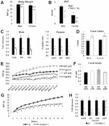
          As compared to wild-type littermates, transgenic hNAG-1 mice have less white fat and brown fat despite equivalent food intake, improved glucose tolerance, lower insulin levels and are resistant to dietary- and genetic-induced obesity. hNAG-1 mice are more metabolically active with higher energy expenditure. Obese C57BL/6 mice treated with hNAG-1 expressing xenografts show decreases in adipose tissue and serum insulin levels. hNAG-1 mice and obese mice treated with hNAG-1 expressing xenografts show increased thermogenic gene expression ( UCP1, PGC1α, ECH1, Cox8b, Dio2, Cyc1, PGC1β, PPARα, Elvol3) in brown adipose tissue (BAT) and increased expression of lipolytic genes ( Adrb3, ATGL, HSL) in both white adipose tissue (WAT) and BAT, consistent with higher energy metabolism

          Conclusion

          hNAG-1 modulates metabolic activity by increasing the expression of key thermogenic and lipolytic genes in BAT and WAT. hNAG-1 appears to be a novel therapeutic target in preventing and treating obesity and insulin resistance.

          Related collections

          Most cited references38

          • Record: found
          • Abstract: found
          • Article: not found

          Adipose tissue dysfunction in obesity, diabetes, and vascular diseases.

          The classical perception of adipose tissue as a storage place of fatty acids has been replaced over the last years by the notion that adipose tissue has a central role in lipid and glucose metabolism and produces a large number of hormones and cytokines, e.g. tumour necrosis factor-alpha, interleukin-6, adiponectin, leptin, and plasminogen activator inhibitor-1. The increased prevalence of excessive visceral obesity and obesity-related cardiovascular risk factors is closely associated with the rising incidence of cardiovascular diseases and type 2 diabetes mellitus. This clustering of vascular risk factors in (visceral) obesity is often referred to as metabolic syndrome. The close relationship between an increased quantity of visceral fat, metabolic disturbances, including low-grade inflammation, and cardiovascular diseases and the unique anatomical relation to the hepatic portal circulation has led to an intense endeavour to unravel the specific endocrine functions of this visceral fat depot. The objective of this paper is to describe adipose tissue dysfunction, delineate the relation between adipose tissue dysfunction and obesity and to describe how adipose tissue dysfunction is involved in the development of diabetes mellitus type 2 and atherosclerotic vascular diseases. First, normal physiology of adipocytes and adipose tissue will be described.
            Bookmark
            • Record: found
            • Abstract: found
            • Article: not found

            Brown adipose tissue oxidative metabolism contributes to energy expenditure during acute cold exposure in humans.

            Brown adipose tissue (BAT) is vital for proper thermogenesis during cold exposure in rodents, but until recently its presence in adult humans and its contribution to human metabolism were thought to be minimal or insignificant. Recent studies using PET with 18F-fluorodeoxyglucose (18FDG) have shown the presence of BAT in adult humans. However, whether BAT contributes to cold-induced nonshivering thermogenesis in humans has not been proven. Using PET with 11C-acetate, 18FDG, and 18F-fluoro-thiaheptadecanoic acid (18FTHA), a fatty acid tracer, we have quantified BAT oxidative metabolism and glucose and nonesterified fatty acid (NEFA) turnover in 6 healthy men under controlled cold exposure conditions. All subjects displayed substantial NEFA and glucose uptake upon cold exposure. Furthermore, we demonstrated cold-induced activation of oxidative metabolism in BAT, but not in adjoining skeletal muscles and subcutaneous adipose tissue. This activation was associated with an increase in total energy expenditure. We found an inverse relationship between BAT activity and shivering. We also observed an increase in BAT radio density upon cold exposure, indicating reduced BAT triglyceride content. In sum, our study provides evidence that BAT acts as a nonshivering thermogenesis effector in humans.
              Bookmark
              • Record: found
              • Abstract: found
              • Article: not found

              New role of bone morphogenetic protein 7 in brown adipogenesis and energy expenditure.

              Adipose tissue is central to the regulation of energy balance. Two functionally different types of fat are present in mammals: white adipose tissue, the primary site of triglyceride storage, and brown adipose tissue, which is specialized in energy expenditure and can counteract obesity. Factors that specify the developmental fate and function of white and brown adipose tissue remain poorly understood. Here we demonstrate that whereas some members of the family of bone morphogenetic proteins (BMPs) support white adipocyte differentiation, BMP7 singularly promotes differentiation of brown preadipocytes even in the absence of the normally required hormonal induction cocktail. BMP7 activates a full program of brown adipogenesis including induction of early regulators of brown fat fate PRDM16 (PR-domain-containing 16; ref. 4) and PGC-1alpha (peroxisome proliferator-activated receptor-gamma (PPARgamma) coactivator-1alpha; ref. 5), increased expression of the brown-fat-defining marker uncoupling protein 1 (UCP1) and adipogenic transcription factors PPARgamma and CCAAT/enhancer-binding proteins (C/EBPs), and induction of mitochondrial biogenesis via p38 mitogen-activated protein (MAP) kinase-(also known as Mapk14) and PGC-1-dependent pathways. Moreover, BMP7 triggers commitment of mesenchymal progenitor cells to a brown adipocyte lineage, and implantation of these cells into nude mice results in development of adipose tissue containing mostly brown adipocytes. Bmp7 knockout embryos show a marked paucity of brown fat and an almost complete absence of UCP1. Adenoviral-mediated expression of BMP7 in mice results in a significant increase in brown, but not white, fat mass and leads to an increase in energy expenditure and a reduction in weight gain. These data reveal an important role of BMP7 in promoting brown adipocyte differentiation and thermogenesis in vivo and in vitro, and provide a potential new therapeutic approach for the treatment of obesity.
                Bookmark

                Author and article information

                Journal
                101256108
                32579
                Int J Obes (Lond)
                Int J Obes (Lond)
                International journal of obesity (2005)
                0307-0565
                1476-5497
                20 March 2014
                17 February 2014
                December 2014
                01 June 2015
                : 38
                : 12
                : 1555-1564
                Affiliations
                [1 ]Laboratory of Molecular Carcinogenesis, NIEHS, NIH, Research Triangle Park, NC 27709
                [2 ]Cellular and Molecular Pathology Branch, NIEHS, NIH, Research Triangle Park, NC 27709
                [3 ]Department of Biomedical and Diagnostic Sciences, College of Veterinary Medicine, University of Tennessee, 2407 River Drive, Knoxville, TN 37996
                Author notes
                []Correspondence: Thomas E. Eling, Laboratory of Molecular Carcinogenesis, 111 T.W. Alexander Drive, Building 101, Room D448B, Research Triangle Park, NC 27709, Phone: 919-541-3911, Fax: 919-541-0146, eling@ 123456niehs.nih.gov
                [4]

                These authors contributed equally to this work

                Article
                NIHMS560855
                10.1038/ijo.2014.27
                4135041
                24531647
                769e2c4b-e876-44c2-bacf-3b68211db3a5
                History
                Categories
                Article

                Nutrition & Dietetics
                nag-1/gdf-15,insulin resistance,bat,thermogenesis,lipolysis
                Nutrition & Dietetics
                nag-1/gdf-15, insulin resistance, bat, thermogenesis, lipolysis

                Comments

                Comment on this article