3
views
0
recommends
+1 Recommend
0 collections
    0
    shares
      • Record: found
      • Abstract: found
      • Article: found
      Is Open Access

      Radiation-induced changes to bone composition extend beyond periosteal bone

      research-article

      Read this article at

      Bookmark
          There is no author summary for this article yet. Authors can add summaries to their articles on ScienceOpen to make them more accessible to a non-specialist audience.

          Abstract

          Background

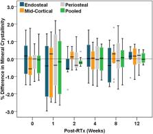
          Cancer patients receiving radiotherapy for soft tissue sarcomas are often at risk of post-irradiation (post-RTx) bone fragility fractures, but our understanding of factors controlling radiation-induced bone injury is limited. Previous studies have evaluated post-RTx changes to cortical bone composition in the periosteum of irradiated tibiae, but have not evaluated effects of irradiation in deeper tissues, such as endosteal or mid-cortical bone, and whether there are differential spatial effects of irradiation. In this study, we hypothesize that post-RTx changes to cortical bone composition are greater in endosteal compared to mid-cortical or periosteal bone.

          Methods

          A pre-clinical mouse model of limited field hindlimb irradiation was used to evaluate spatial and temporal post-RTx changes to the metaphyseal cortex of irradiated tibiae. Irradiation was delivered unilaterally to the hindlimbs of 12-wk old female BALB/cJ mice as 4 consecutive daily doses of 5 Gy each. RTx and non-RTx tibiae were obtained at 0, 2, 4, 8, and 12 wks post-RTx ( n = 9 mice/group/time). Raman spectroscopy was used to evaluate spatial and temporal post-RTx changes to cortical bone composition in age-matched RTx and non-RTx groups.

          Results

          Significant early spatial differences in mineral/matrix and collagen crosslink ratios were found between endosteal and periosteal or mid-cortical bone at 2-wks post-RTx. Although spatial differences were transient, mineral/matrix ratios significantly decreased and collagen crosslink ratios significantly increased with post-RTx time throughout the entire tibial metaphyseal cortex.

          Conclusions

          Irradiation negatively impacts the composition of cortical bone in a spatially-dependent manner starting as early as 2-wks post-RTx. Long-term progressive post-RTx changes across all cortical bone sites may eventually contribute to the increased risk of post-RTx bone fragility fractures.

          Related collections

          Most cited references61

          • Record: found
          • Abstract: found
          • Article: not found

          Spectroscopic characterization of collagen cross-links in bone.

          Collagen is the most abundant protein of the organic matrix in mineralizing tissues. One of its most critical properties is its cross-linking pattern. The intermolecular cross-linking provides the fibrillar matrices with mechanical properties such as tensile strength and viscoelasticity. In this study, Fourier transform infrared (FTIR) spectroscopy and FTIR imaging (FTIRI) analyses were performed in a series of biochemically characterized samples including purified collagen cross-linked peptides, demineralized bovine bone collagen from animals of different ages, collagen from vitamin B6-deficient chick homogenized bone and their age- and sex-matched controls, and histologically stained thin sections from normal human iliac crest biopsy specimens. One region of the FTIR spectrum of particular interest (the amide I spectral region) was resolved into its underlying components. Of these components, the relative percent area ratio of two subbands at approximately 1660 cm(-1) and approximately 1690 cm(-1) was related to collagen cross-links that are abundant in mineralized tissues (i.e., pyridinoline [Pyr] and dehydrodihydroxylysinonorleucine [deH-DHLNL]). This study shows that it is feasible to monitor Pyr and DHLNL collagen cross-links spatial distribution in mineralized tissues. The spectroscopic parameter established in this study may be used in FTIRI analyses, thus enabling the calculation of relative Pyr/DHLNL amounts in thin (approximately 5 microm) calcified tissue sections with a spatial resolution of approximately 7 microm.
            Bookmark
            • Record: found
            • Abstract: found
            • Article: not found

            Carbonate assignment and calibration in the Raman spectrum of apatite.

            A series of apatites with varying carbonate levels was prepared in order to assign the carbonate bands and calibrate for Raman analysis of natural materials. Overlap of carbonate bands with phosphate peaks was resolved by curve fitting. A peak at 1,071 cm(-1) was assigned to a combination of the carbonate nu(1) mode at 1,070 cm(-1) with a phosphate nu(3) mode at 1,076 cm(-1). In addition, the carbonate nu(4) mode was identified in apatite samples with >4% carbonate. The carbonate nu(4) bands at 715 and 689 cm(-1) identify the samples as B-type carbonated apatite. The carbonate content of apatite was calibrated to a carbonate Raman band, and the method was used to determine the carbonate content of a sample of bovine cortical bone, 7.7 +/- 0.4%.
              Bookmark
              • Record: found
              • Abstract: found
              • Article: not found

              Risk of pelvic fractures in older women following pelvic irradiation.

              Pelvic fractures, including hip fractures, are a major source of morbidity and mortality in older women. Although therapeutic pelvic irradiation could increase the risk of such fractures, this effect has not been studied. To determine if women who undergo pelvic irradiation for pelvic malignancies (anal, cervical, or rectal cancers) have a higher rate of pelvic fracture than women with pelvic malignancies who do not undergo irradiation. We conducted a retrospective cohort study using Surveillance, Epidemiology, and End Results (SEER) cancer registry data linked to Medicare claims data. A total of 6428 women aged 65 years and older diagnosed with pelvic malignancies from 1986 through 1999 were included. We compared results for women who did (n = 2855) vs did not (n = 3573) undergo radiation therapy. To assess the influence of selection bias, we also evaluated the effect of irradiation on osteoporotic fractures in nonirradiated sites (arm and spine). We evaluated the effect of irradiation on the incidence of pelvic fractures over time, and adjusted for potential confounders using a proportional hazards model. Women who underwent radiation therapy were more likely to have a pelvic fracture than women who did not undergo radiation therapy (cumulative 5-year fracture rate, 14.0% vs 7.5% in women with anal cancer, 8.2% vs 5.9% in women with cervical cancer, and 11.2% vs 8.7% in women with rectal cancer); the difference was statistically significant and most fractures (90%) were hip fractures. We controlled for potential confounders including age, race, cancer stage, and geographic location. The impact of irradiation varied by cancer site: treatment for anal cancer was associated with a higher risk of pelvic fractures (hazard ratio, 3.16; 95% confidence interval, 1.48-6.73); than for cervical cancer (hazard ratio, 1.66; 95% confidence interval, 1.06-2.59); or rectal cancer (hazard ratio, 1.65; 95% confidence interval, 1.33-2.05). No statistically significant difference was found in the rate of arm or spine fractures between the irradiated and nonirradiated groups (hazard ratio, 1.15; 95% confidence interval, 0.89-1.48). Pelvic irradiation substantially increases the risk of pelvic fractures in older women. Given the high baseline risk of pelvic fracture, this finding is of particular concern.
                Bookmark

                Author and article information

                Contributors
                Journal
                Bone Rep
                Bone Rep
                Bone Reports
                Elsevier
                2352-1872
                28 March 2020
                June 2020
                28 March 2020
                : 12
                : 100262
                Affiliations
                [a ]School of Dentistry, University of Michigan, Ann Arbor, MI, USA
                [b ]Department of Orthopedic Surgery, Upstate Medical University, Syracuse, NY, USA
                [c ]Department of Chemistry, University of Michigan, Ann Arbor, USA
                [d ]Department of Biomedical Engineering, University of Michigan, Ann Arbor, USA
                Author notes
                [* ]Corresponding author at: Departments of Biologic and Materials Sciences and Biomedical Engineering, University of Michigan, Ann Arbor, MI 48109-1078, USA. dhkohn@ 123456umich.edu
                Article
                S2352-1872(20)30022-X 100262
                10.1016/j.bonr.2020.100262
                7125315
                32258252
                76529a91-efc6-46ef-9cc8-bb0b577bdd05
                © 2020 The Author(s)

                This is an open access article under the CC BY-NC-ND license (http://creativecommons.org/licenses/by-nc-nd/4.0/).

                History
                : 8 December 2019
                : 25 March 2020
                : 26 March 2020
                Categories
                Article

                post-irradiation,mouse model,raman spectroscopy,bone quality,cortical bone,bone composition

                Comments

                Comment on this article