11
views
0
recommends
+1 Recommend
0 collections
    0
    shares
      • Record: found
      • Abstract: found
      • Article: found
      Is Open Access

      Exosomes derived from human adipose mesenchymal stem cells improve ovary function of premature ovarian insufficiency by targeting SMAD

      research-article

      Read this article at

      Bookmark
          There is no author summary for this article yet. Authors can add summaries to their articles on ScienceOpen to make them more accessible to a non-specialist audience.

          Abstract

          Background

          Although many reports show that various kinds of stem cells have the ability to recover the function of premature ovarian insufficiency (POI), few studies are associated with the mechanism of stem cell treatment of POI. We designed this experimental study to investigate whether human adipose stem cell-derived exosomes (hADSC-Exos) retain the ability to restore ovarian function and how hADSC-Exos work in this process.

          Methods

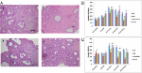
          A POI mouse model was established and human ovarian granule cells (hGCs) collected from individuals with POI were prepared to assess the therapeutic effects and illuminate the mechanism of hADSCs in curing POI. The hematoxylin and eosin assay method was employed to assess the number of follicles. Enzyme-linked immunosorbent assay (ELISA) was used to detect the serum levels of sex hormones. The proliferation rate and marker expression levels of hGCs were measured by flow cytometry (fluorescence-activated cell sorting). Real-time PCR and western blot assays were used to determine the mRNA and protein expression levels of SMAD2, SMAD3, and SMAD5. Western blot assays were used to test the protein expression levels of apoptosis genes (Fas, FasL, caspase-3, and caspase-8).

          Results

          After the hADSC-Exos were transplanted into the POI mice model, they exerted better therapeutic activity on mouse ovarian function, improving follicle numbers during four stages. ELISA results showed that hADSC-Exos elevated the hormone levels to the normal levels. In addition, after hADSC-Exos were cocultured with POI hGCs, our results showed that hADSC-Exos significantly promoted the proliferation rate and inhibited the apoptosis rate. Furthermore, hADSC-Exos also increased the marker expression of hGCs to the normal level. Besides, mRNA and protein assays demonstrated that hADSC-Exos downregulated the expression of SMAD2, SMAD3, and SMAD5 in vivo and in vitro. Western blot assay demonstrated that hADSC-Exos inhibited expression of the apoptosis genes in POI hGCs, and SMAD knockdown increased the protein expression of apoptosis genes.

          Conclusions

          These findings demonstrate for the first time the molecular cascade and related cell biology events involved in the mechanism by which exosomes derived from hADSCs improved ovarian function of POI disease via regulation of the SMAD signaling pathway.

          Electronic supplementary material

          The online version of this article (10.1186/s13287-018-0953-7) contains supplementary material, which is available to authorized users.

          Related collections

          Most cited references19

          • Record: found
          • Abstract: found
          • Article: found
          Is Open Access

          Exosomes derived from human menstrual blood-derived stem cells alleviate fulminant hepatic failure

          Background Human menstrual blood-derived stem cells (MenSCs) are a novel source of MSCs that provide the advantage of being easy to collect and isolate. Exosomes contain some mRNAs and adhesion molecules that can potentially impact cellular and animal physiology. This study aimed to investigate the therapeutic potential of MenSC-derived exosomes (MenSC-Ex) on AML12 cells (in vitro) and D-GalN/LPS-induced FHF mice (in vivo). Methods Transmission electron microscopy and Western blot were used to identify MenSC-Ex. Antibody array was used to examine cytokine levels on MenSC-Ex. MenSC-Ex were treated in D-GalN/LPS-induced AML12 in vitro. Cell proliferation and apoptosis were measured. MenSC-Ex were injected into the tail veins of mice 24 h before treatment with D-GalN/LPS. Blood and liver tissues served as physiological and biochemical indexes. The number of liver mononuclear cells (MNCs) and the amount of the active apoptotic protein caspase-3 were determined to elaborate the mechanism of hepatoprotective activity. Results Human menstrual blood-derived stem cell-derived exosomes (MenSC-Ex) are bi-lipid membrane vesicles that have a round, ball-like shape with a diameter of approximately 30–100 nm. Cytokine arrays have shown that MenSC-Ex expressed cytokines, including ICAM-1, angiopoietin-2, Axl, angiogenin, IGFBP-6, osteoprotegerin, IL-6, and IL-8. MenSC-Ex markedly improved liver function, enhanced survival rates, and inhibited liver cell apoptosis at 6 h after transplantation. MenSC-Ex migrated to sites of injury and to AML12 cells (a mouse hepatocyte cell line), respectively. Moreover, MenSC-Ex reduced the number of liver mononuclear cells (MNCs) and the amount of the active apoptotic protein caspase-3 in injured livers. Conclusions In conclusion, our results provide preliminary evidence for the anti-apoptotic capacity of MenSC-Ex in FHF and suggest that MenSC-Ex may be an alternative therapeutic approach to treat FHF. Electronic supplementary material The online version of this article (doi:10.1186/s13287-016-0453-6) contains supplementary material, which is available to authorized users.
            Bookmark
            • Record: found
            • Abstract: found
            • Article: found
            Is Open Access

            Adipose-derived stem cells improved mouse ovary function after chemotherapy-induced ovary failure

            Introduction Young patients receiving chemotherapy occasionally face infertility and premature ovarian failure (POF). Numerous investigations reported that adipose-derived stem cells (ADSCs) transplantation could ameliorate the structure and function of injured tissues. The aim of this study was to explore the therapeutic efficacy of ADSC transplantation for chemotherapy-induced ovarian damage. Methods Female mice were injected intraperitoneally with 50 mg/kg cyclophosphamide (CTX). After 15 consecutive days of injection, ADSCs were transplanted either directly into bilateral ovaries or via intravenous injection, and the ovaries were excised after either 1 week or 1 month of treatment. The follicles were counted and categorized, and ovarian histologic sections were stained for TUNEL. Ovarian function was evaluated by monitoring ovulation. ADSC tracking, microarray analyses, and real-time polymerase chain reaction (PCR) were used to assess the inner mechanism of injury and repair. Results The ovarian function of mice exposed to CTX injection improved after ADSC transplantation. The population of follicles at different stages and ovulation significantly increased after the treatment. Immunofluorescence revealed reduced TUNEL staining. The tracking of ADSCs revealed that these cells did not directly differentiate into the follicle component. Microarray analyses indicated that changes in different groups of genes might affect follicle formation or ovulation. Conclusions ADSC transplantation improved ovarian function. Our results suggest a potential mechanism for ADSC therapy.
              Bookmark
              • Record: found
              • Abstract: found
              • Article: found
              Is Open Access

              The restorative effects of adipose-derived mesenchymal stem cells on damaged ovarian function

              The clinical application of human adipose-derived mesenchymal stem cells (MSCs) as treatment for intractable diseases or traumatic tissue damage has attracted attention. To address the ability of reactivating injured ovaries, we prepared a rat model with damaged ovaries by using an anticancer agent, cyclophosphamide (CTX). We then investigated the restorative effects on ovarian function and the safety of adipose-derived MSCs (A-MSCs). MSCs were shown to be capable of inducing angiogenesis and restoring the number of ovarian follicles and corpus lutea in ovaries. No deformities, tumor formation or deaths were observed in F1 and F2 rats, indicating that the local injection of MSCs into the ovary did not have any obvious side effects. In addition, the localization of the Y chromosome was investigated using the fluorescent in situ hybridization method by injecting male A-MSCs into the ovaries; as a result, the Y chromosomes were localized not in the follicles, but in the thecal layers. ELISA revealed that A-MSCs secreted higher levels of vascular endothelial cell growth factor (VEGF), insulin-like growth factor-1 (IGF-1) and hepatocyte growth factor (HGF) than tail fibroblast cells. Quantitative real-time PCR and immunohistochemistry showed that higher expression levels of VEGF, IGF-1 and HGF were observed in CTX-treated ovaries after A-MSC transplantation. These findings suggest that MSCs may have a role in restoring damaged ovarian function and could be useful for regenerative medicine.
                Bookmark

                Author and article information

                Contributors
                +86-512-62362461 , huangboxiannj@163.com
                lujiafeng1984@163.com
                dingchenyue89@163.com
                zouqinyan1975@163.com
                157304321@qq.com
                +86-512-62362461 , hongliszivf@163.com
                Journal
                Stem Cell Res Ther
                Stem Cell Res Ther
                Stem Cell Research & Therapy
                BioMed Central (London )
                1757-6512
                9 August 2018
                9 August 2018
                2018
                : 9
                : 216
                Affiliations
                [1 ]GRID grid.440227.7, Center of Reproduction and Genetics, , Affiliated Suzhou Hospital of Nanjing Medical University, Suzhou Municipal Hospital, ; Suzhou, 215002 China
                [2 ]GRID grid.440227.7, Central Laboratory, , Affiliated Suzhou Hospital of Nanjing Medical University, Suzhou Municipal Hospital, ; Suzhou, 215002 China
                [3 ]ISNI 0000 0000 9255 8984, GRID grid.89957.3a, State Key Laboratory of Reproductive Medicine, , Nanjing Medical University, ; Nanjing, 210029 China
                Author information
                http://orcid.org/0000-0001-9913-9077
                Article
                953
                10.1186/s13287-018-0953-7
                6085638
                30092819
                75d138b5-96e1-4e9e-b892-3c2eeea243db
                © The Author(s). 2018

                Open AccessThis article is distributed under the terms of the Creative Commons Attribution 4.0 International License ( http://creativecommons.org/licenses/by/4.0/), which permits unrestricted use, distribution, and reproduction in any medium, provided you give appropriate credit to the original author(s) and the source, provide a link to the Creative Commons license, and indicate if changes were made. The Creative Commons Public Domain Dedication waiver ( http://creativecommons.org/publicdomain/zero/1.0/) applies to the data made available in this article, unless otherwise stated.

                History
                : 1 May 2018
                : 26 June 2018
                : 6 July 2018
                Funding
                Funded by: Suzhou introduce expert team of clinical medicine
                Award ID: SZYJTD201708
                Award Recipient :
                Funded by: Suzhou key medicine center
                Award ID: SZZX201505
                Award Recipient :
                Funded by: Jiangsu Provincial medical innovation team
                Award ID: CXTDB2017013
                Award Recipient :
                Funded by: Maternal and health care project of Jiangsu Province-assisted reproduction medicine center
                Award ID: FKX201224
                Award Recipient :
                Categories
                Research
                Custom metadata
                © The Author(s) 2018

                Molecular medicine
                human adipose stem cells,premature ovarian insufficiency,exosome,smad pathway

                Comments

                Comment on this article