2
views
0
recommends
+1 Recommend
0 collections
    0
    shares
      • Record: found
      • Abstract: found
      • Article: found
      Is Open Access

      The methionine salvage pathway-involving ADI1 inhibits hepatoma growth by epigenetically altering genes expression via elevating S-adenosylmethionine

      research-article

      Read this article at

      Bookmark
          There is no author summary for this article yet. Authors can add summaries to their articles on ScienceOpen to make them more accessible to a non-specialist audience.

          Abstract

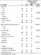
          The 5′-methylthioadenosine (MTA) cycle-participating human acireductone dioxygenase 1 (ADI1) has been implicated as a tumor suppressor in prostate cancer, yet its role remains unclear in hepatocellular carcinoma (HCC). Here, we demonstrated a significant reduction of ADI1, either in protein or mRNA level, in HCC tissues. Additionally, higher ADI1 levels were associated with favorable postoperative recurrence-free survival in HCC patients. By altering ADI1 expression in HCC cells, a negative correlation between ADI1 and cell proliferation was observed. Cell-based and xenograft experiments were performed by using cells overexpressing ADI1 mutants carrying mutations at the metal-binding sites (E94A and H133A, respectively), which selectively disrupted differential catalytic steps, resulting in staying or leaving the MTA cycle. The results showed that the growth suppression effect was mediated by accelerating the MTA cycle. A cDNA microarray analysis followed by verification experiments identified that caveolin-1 (CAV1), a growth-promoting protein in HCC, was markedly decreased upon ADI1 overexpression. Suppression of CAV1 expression was mediated by an increase of S-adenosylmethionine (SAMe) level. The methylation status of CAV1 promoter was significantly altered upon ADI1 overexpression. Finally, a genome-wide methylation analysis revealed that ADI1 overexpression altered promoter methylation profiles in a set of cancer-related genes, including CAV1 and genes encoding antisense non-coding RNAs, long non-coding RNAs, and microRNAs, resulting in significant changes of their expression levels. In conclusion, ADI1 expression promoted MTA cycle to increase SAMe levels, which altered genome-wide promoter methylation profiles, resulting in altered gene expression and HCC growth suppression.

          Related collections

          Most cited references41

          • Record: found
          • Abstract: found
          • Article: found
          Is Open Access

          Long Intergenic Noncoding RNA 00511 Acts as an Oncogene in Non–small-cell Lung Cancer by Binding to EZH2 and Suppressing p57

          Long noncoding RNAs (lncRNAs) play crucial roles in carcinogenesis. However, the function and mechanism of lncRNAs in human non–small-cell lung cancer (NSCLC) are still remaining largely unknown. Long intergenic noncoding RNA 00511 (LINC00511) has been found to be upregulated and acts as an oncogene in breast cancer, but little is known about its expression pattern, biological function and underlying mechanism in NSCLC. Herein, we identified LINC00511 as an oncogenic lncRNA by driving tumorigenesis in NSCLC. We found LINC00511 was upregulated and associated with oncogenesis, tumor size, metastasis, and poor prognosis in NSCLC. Moreover, LINC00511 affected cell proliferation, invasiveness, metastasis, and apoptosis in multiple NSCLC cell lines. Mechanistically, LINC00511 bound histone methyltransferase enhancer of zeste homolog 2 ((EZH2, the catalytic subunit of the polycomb repressive complex 2 (PRC2), a highly conserved protein complex that regulates gene expression by methylating lysine 27 on histone H3), and acted as a modular scaffold of EZH2/PRC2 complexes, coordinated their localization, and specified the histone modification pattern on the target genes, including p57, and consequently altered NSCLC cell biology. Thus, LINC00511 is mechanistically, functionally, and clinically oncogenic in NSCLC. Targeting LINC00511 and its pathway may be meaningful for treating patients with NSCLC.
            Bookmark
            • Record: found
            • Abstract: found
            • Article: not found

            A new class of quinoline-based DNA hypomethylating agents reactivates tumor suppressor genes by blocking DNA methyltransferase 1 activity and inducing its degradation.

            Reactivation of silenced tumor suppressor genes by 5-azacytidine (Vidaza) and its congener 5-aza-2'-deoxycytidine (decitabine) has provided an alternate approach to cancer therapy. We have shown previously that these drugs selectively and rapidly induce degradation of the maintenance DNA methyltransferase (DNMT) 1 by a proteasomal pathway. Because the toxicity of these compounds is largely due to their incorporation into DNA, it is critical to explore novel, nonnucleoside compounds that can effectively reactivate the silenced genes. Here, we report that a quinoline-based compound, designated SGI-1027, inhibits the activity of DNMT1, DNMT3A, and DNMT3B as well M. SssI with comparable IC(50) (6-13 micromol/L) by competing with S-adenosylmethionine in the methylation reaction. Treatment of different cancer cell lines with SGI-1027 resulted in selective degradation of DNMT1 with minimal or no effects on DNMT3A and DNMT3B. At a concentration of 2.5 to 5 micromol/L (similar to that of decitabine), complete degradation of DNMT1 protein was achieved within 24 h without significantly affecting its mRNA level. MG132 blocked SGI-1027-induced depletion of DNMT1, indicating the involvement of proteasomal pathway. Prolonged treatment of RKO cells with SGI-1027 led to demethylation and reexpression of the silenced tumor suppressor genes P16, MLH1, and TIMP3. Further, this compound did not exhibit significant toxicity in a rat hepatoma (H4IIE) cell line. This study provides a novel class of DNA hypomethylating agents that have the potential for use in epigenetic cancer therapy.
              Bookmark
              • Record: found
              • Abstract: found
              • Article: not found
              Is Open Access

              Pleiotropic effects of methionine adenosyltransferases deregulation as determinants of liver cancer progression and prognosis.

              Downregulation of liver-specific MAT1A gene, encoding S-adenosylmethionine (SAM) synthesizing isozymes MATI/III, and upregulation of widely expressed MAT2A, encoding MATII isozyme, known as MAT1A:MAT2A switch, occurs in hepatocellular carcinoma (HCC). Being inhibited by its reaction product, MATII isoform upregulation cannot compensate for MATI/III decrease. Therefore, MAT1A:MAT2A switch contributes to decrease in SAM level in rodent and human hepatocarcinogenesis. SAM administration to carcinogen-treated rats prevents hepatocarcinogenesis, whereas MAT1A-KO mice, characterized by chronic SAM deficiency, exhibit macrovesicular steatosis, mononuclear cell infiltration in periportal areas, and HCC development. This review focuses upon the pleiotropic changes, induced by MAT1A/MAT2A switch, associated with HCC development. Epigenetic control of MATs expression occurs at transcriptional and post-transcriptional levels. In HCC cells, MAT1A/MAT2A switch is associated with global DNA hypomethylation, decrease in DNA repair, genomic instability, and signaling deregulation including c-MYC overexpression, rise in polyamine synthesis, upregulation of RAS/ERK, IKK/NF-kB, PI3K/AKT, and LKB1/AMPK axis. Furthermore, decrease in MAT1A expression and SAM levels results in increased HCC cell proliferation, cell survival, and microvascularization. All of these changes are reversed by SAM treatment in vivo or forced MAT1A overexpression or MAT2A inhibition in cultured HCC cells. In human HCC, MAT1A:MAT2A and MATI/III:MATII ratios correlate negatively with cell proliferation and genomic instability, and positively with apoptosis and global DNA methylation. This suggests that SAM decrease and MATs deregulation represent potential therapeutic targets for HCC. Finally, MATI/III:MATII ratio strongly predicts patients' survival length suggesting that MAT1A:MAT2A expression ratio is a putative prognostic marker for human HCC. Copyright © 2013 European Association for the Study of the Liver. Published by Elsevier B.V. All rights reserved.
                Bookmark

                Author and article information

                Contributors
                +886-3-3281200 , chautingy@gmail.com
                Journal
                Cell Death Dis
                Cell Death Dis
                Cell Death & Disease
                Nature Publishing Group UK (London )
                2041-4889
                11 March 2019
                11 March 2019
                March 2019
                : 10
                : 3
                : 240
                Affiliations
                [1 ]ISNI 0000 0001 0711 0593, GRID grid.413801.f, Liver Research Center, , Chang Gung Memorial Hospital, ; Taoyuan, Taiwan
                [2 ]GRID grid.145695.a, Molecular Medicine Research Center, , Chang Gung University, ; Taoyuan, Taiwan
                [3 ]GRID grid.145695.a, Department of Biochemistry, , Chang Gung University, ; Taoyuan City, Taiwan
                [4 ]GRID grid.145695.a, Graduate Institute of Biomedical Sciences, College of Medicine, , Chang Gung University, ; Taoyuan City, Taiwan
                [5 ]ISNI 0000 0004 0604 5314, GRID grid.278247.c, Medical Research Department, , Taipei Veterans General Hospital, ; Taipei, Taiwan
                Article
                1486
                10.1038/s41419-019-1486-4
                6411897
                30858354
                75797a5a-c244-4e8c-9220-6e28aa20039a
                © The Author(s) 2019

                Open Access This article is licensed under a Creative Commons Attribution 4.0 International License, which permits use, sharing, adaptation, distribution and reproduction in any medium or format, as long as you give appropriate credit to the original author(s) and the source, provide a link to the Creative Commons license, and indicate if changes were made. The images or other third party material in this article are included in the article’s Creative Commons license, unless indicated otherwise in a credit line to the material. If material is not included in the article’s Creative Commons license and your intended use is not permitted by statutory regulation or exceeds the permitted use, you will need to obtain permission directly from the copyright holder. To view a copy of this license, visit http://creativecommons.org/licenses/by/4.0/.

                History
                : 1 November 2018
                : 18 February 2019
                : 25 February 2019
                Funding
                Funded by: FundRef https://doi.org/10.13039/501100005795, Chang Gung Memorial Hospital, Linkou (Linkou Chang Gung Memorial Hospital);
                Award ID: CMRPG3F1602
                Award Recipient :
                Categories
                Article
                Custom metadata
                © The Author(s) 2019

                Cell biology
                Cell biology

                Comments

                Comment on this article