15
views
0
recommends
+1 Recommend
0 collections
    0
    shares
      • Record: found
      • Abstract: found
      • Article: found
      Is Open Access

      Protein losing enteropathy: comprehensive review of the mechanistic association with clinical and subclinical disease states

      review-article

      Read this article at

      Bookmark
          There is no author summary for this article yet. Authors can add summaries to their articles on ScienceOpen to make them more accessible to a non-specialist audience.

          Abstract

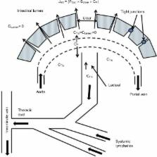
          Protein losing enteropathy (PLE) has been associated with more than 60 different conditions, including nearly all gastrointestinal diseases (Crohn’s disease, celiac, Whipple’s, intestinal infections, and so on) and a large number of non-gut conditions (cardiac and liver disease, lupus, sarcoidosis, and so on). This review presents the first attempt to quantitatively understand the magnitude of the PLE in relation to the associated pathology for three different disease categories: 1) increased lymphatic pressure (e.g., lymphangiectasis); 2) diseases with mucosal erosions (e.g., Crohn’s disease); and 3) diseases without mucosal erosions (e.g., celiac disease). The PLE with lymphangiectasis results from rupture of the mucosal lymphatics, with retrograde drainage of systemic lymph into the intestinal lumen with the resultant loss of CD4 T cells, which is diagnostic. Mucosal erosion PLE results from macroscopic breakdown of the mucosal barrier, with the epithelial capillaries becoming the rate-limiting factor in albumin loss. The equation derived to describe the relationship between the reduction in serum albumin (C P) and PLE indicates that gastrointestinal albumin clearance must increase by at least 17 times normal to reduce the C P by half. The strengths and limitations of the two quantitative measures of PLE ( 51Cr-albumin or α 1-antitrypsin [αAT] clearance) are reviewed. αAT provides a simple quantitative diagnostic test that is probably underused clinically. The strong, unexplained correlation between minor decreases in C P and subsequent mortality in seemingly healthy individuals raises the question of whether subclinical PLE could account for the decreased C P and, if so, could the mechanism responsible for PLE play a role in the increased mortality? A large-scale study correlating αAT clearance with serum albumin concentrations will be required in order to determine the role of PLE in the regulation of the serum albumin concentration of seemingly healthy subjects.

          Related collections

          Most cited references123

          • Record: found
          • Abstract: found
          • Article: not found

          Loss of Infliximab Into Feces Is Associated With Lack of Response to Therapy in Patients With Severe Ulcerative Colitis.

          It is not clear why some patients with ulcerative colitis (UC) do not respond to treatment with anti-tumor necrosis factor (TNF) agents, such as infliximab. It could be that some patients have high level of inflammation, with large quantities of TNF to be neutralized by the drug. We investigated whether loss of anti-TNF agents through ulcerated intestinal mucosa reduces the efficacy of these drugs in patients with severe UC.
            Bookmark
            • Record: found
            • Abstract: found
            • Article: not found

            MYO5B mutations cause microvillus inclusion disease and disrupt epithelial cell polarity.

            Following homozygosity mapping in a single kindred, we identified nonsense and missense mutations in MYO5B, encoding type Vb myosin motor protein, in individuals with microvillus inclusion disease (MVID). MVID is characterized by lack of microvilli on the surface of enterocytes and occurrence of intracellular vacuolar structures containing microvilli. In addition, mislocalization of transferrin receptor in MVID enterocytes suggests that MYO5B deficiency causes defective trafficking of apical and basolateral proteins in MVID.
              Bookmark
              • Record: found
              • Abstract: found
              • Article: not found

              A porous defense: the leaky epithelial barrier in intestinal disease.

              A critical function of the intestinal mucosa is to form a barrier that separates luminal contents from the interstitium. This intestinal barrier is compromised in a number of intestinal diseases, most notably inflammatory bowel disease. In vitro studies have demonstrated that cytokines elaborated by immune cells can cause the mucosal barrier to become leaky; these cytokines are known to be increased in intestinal mucosa involved in inflammatory bowel disease. Detailed information describing the mechanisms by which altered cytokine signaling occurs is not available, but recent data implicate the cytoskeleton within epithelial cells as a critical regulator of the mucosal barrier under physiological and pathophysiological conditions. Using available data, we describe a model of intestinal disease where an initial insult to the epithelial barrier may trigger a self-amplifying cycle of immune activation, cytokine release, and further barrier dysfunction. This model is supported by the observation that pharmacological abrogation of cytokine signaling corrects both barrier defects and clinical disease in animal models and human patients, although such therapy clearly has multiple mechanisms. Other therapeutic targets that represent strategies to prevent or reverse disease processes are also considered. The overarching hypothesis is that modulation of the mucosal epithelial barrier plays a critical role in the initiation and propogation of inflammatory intestinal diseases.
                Bookmark

                Author and article information

                Journal
                Clin Exp Gastroenterol
                Clin Exp Gastroenterol
                Clinical and Experimental Gastroenterology
                Clinical and Experimental Gastroenterology
                Dove Medical Press
                1178-7023
                2017
                17 July 2017
                : 10
                : 147-168
                Affiliations
                [1 ]Department of Integrative Biology and Physiology, University of Minnesota
                [2 ]Research Service, Veterans Affairs Medical Center, Minneapolis, MN, USA
                Author notes
                Correspondence: David G Levitt, Department of Integrative Biology and Physiology, University of Minnesota, 6-125 Jackson Hall, 321 Church St S. E., Minneapolis, MN 55455, USA, Tel +1 612 625 7649, Fax +1 612 625 5149, Email levit001@ 123456umn.edu
                [*]

                These authors contributed equally to this work

                Article
                ceg-10-147
                10.2147/CEG.S136803
                5522668
                28761367
                745e10ec-cc06-4b25-85ad-7b82f7f2539d
                © 2017 Levitt and Levitt. This work is published and licensed by Dove Medical Press Limited

                The full terms of this license are available at https://www.dovepress.com/terms.php and incorporate the Creative Commons Attribution – Non Commercial (unported, v3.0) License ( http://creativecommons.org/licenses/by-nc/3.0/). By accessing the work you hereby accept the Terms. Non-commercial uses of the work are permitted without any further permission from Dove Medical Press Limited, provided the work is properly attributed.

                History
                Categories
                Review

                Gastroenterology & Hepatology
                enteropathy,albumin,crohn’s,celiac,lymphangiectasis,antitrypsin
                Gastroenterology & Hepatology
                enteropathy, albumin, crohn’s, celiac, lymphangiectasis, antitrypsin

                Comments

                Comment on this article