12
views
0
recommends
+1 Recommend
0 collections
    0
    shares
      • Record: found
      • Abstract: found
      • Article: found
      Is Open Access

      Bone marrow metastasis presenting as bicytopenia originating from hepatocellular carcinoma

      case-report

      Read this article at
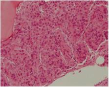
      Bookmark
          There is no author summary for this article yet. Authors can add summaries to their articles on ScienceOpen to make them more accessible to a non-specialist audience.

          Abstract

          The bone is a common site for metastasis in hepatocellular carcinoma (HCC). However, bone marrow metastasis from HCC is rarely reported, and its frequency is unclear. Here we report a rare case of bone marrow metastasis that presented as bicytopenia originating from HCC without bone metastasis. A 58-year-old man was admitted for investigation of a liver mass with extensive lymph node enlargement that was detected when examining his general weakness and weight loss. Laboratory findings revealed anemia, thrombocytopenia, mild elevated liver enzymes, normal prothrombin time percentage and high levels of tumor markers (α-fetoprotein and des-γ-carboxyprothrombin). Abdominal computed tomography showed multiple enhanced masses in the liver and multiple enlarged lymph nodes in the abdomen. A bone marrow biopsy revealed only a few normal hematopoietic cells and abundant tumor cells. Despite its rarity, bone marrow metastasis should always be suspected in HCC patients even if accompanied by cirrhosis.

          Related collections

          Most cited references15

          • Record: found
          • Abstract: found
          • Article: not found

          Extrahepatic metastases of hepatocellular carcinoma.

          To determine the relative frequency, incidence, and locations of metastases of hepatocellular carcinoma (HCC), correlate extrahepatic metastatic disease with intrahepatic tumor staging, and determine the computed tomographic (CT) manifestations of HCC metastases. CT findings in 403 consecutive patients with HCC at our institution since 1992 were reviewed retrospectively. One hundred forty-eight patients with extrahepatic metastatic HCC were identified, and the locations, sizes, and attenuation and enhancement characteristics of the lesions were recorded. A majority (128 [86%] of 148) of patients with extrahepatic HCC foci had either intrahepatic stage IVA tumor (112 [76%] patients) or an intrahepatic stage III tumor (16 [11%] patients) at the occurrence of metastases. The most frequent site of the first detectable metastasis was the lung (58 [39%] patients). Tabulation of all extrahepatic metastatic sites showed the most common to be the lung in 81 (55%) patients, the abdominal lymph nodes in 60 (41%) patients, and the bone in 41 (28%) patients. The lung, abdominal lymph nodes, and bone are the most common sites of extrahepatic metastatic HCC. Most extrahepatic HCC occurs in patients with advanced intrahepatic tumor stage (stage IVA). Incidental extrahepatic lesions at CT in patients with stage I or II intrahepatic HCC are unlikely to represent metastatic HCC.
            Bookmark
            • Record: found
            • Abstract: found
            • Article: not found

            Clinical features and prognosis of patients with extrahepatic metastases from hepatocellular carcinoma.

            To assess the clinical features and prognosis of 151 patients with extrahepatic metastases from primary hepatocellular carcinoma (HCC), and describe the treatment strategy for such patients. After the diagnosis of HCC, all 995 consecutive HCC patients were followed up at regular intervals and 151 (15.2%) patients were found to have extrahepatic metastases at the initial diagnosis of primary HCC or developed such tumors during the follow-up period. We assessed their clinical features, prognosis, and treatment strategies. The most frequent site of extrahepatic metastases was the lungs (47%), followed by lymph nodes (45%), bones (37%), and adrenal glands (12%). The cumulative survival rates after the initial diagnosis of extrahepatic metastases at 6, 12, 24, and 36 mo were 44.1%, 21.7%, 14.2%, 7.1%, respectively. The median survival time was 4.9 mo (range, 0-37 mo). Fourteen patients (11%) died of extrahepatic HCC, others died of primary HCC or liver failure. The prognosis of HCC patients with extrahepatic metastases is poor. With regard to the cause of death, many patients would die of intrahepatic HCC and few of extrahepatic metastases. Although most of HCC patients with extrahepatic metastases should undergo treatment for the primary HCC mainly, treatment of extrahepatic metastases in selected HCC patients who have good hepatic reserve, intrahepatic tumor stage (T0-T2), and are free of portal venous invasion may improve survival.
              Bookmark
              • Record: found
              • Abstract: found
              • Article: not found

              Results of surgical and nonsurgical treatment for small-sized hepatocellular carcinomas: a retrospective and nationwide survey in Japan. The Liver Cancer Study Group of Japan.

              Hepatic resection (HX), percutaneous ethanol injection (PEI), and transcatheter arterial embolization (TCAE) have all been used in the treatment of patients with small-sized hepatocellular carcinomas (HCCs). However, the indications for these therapeutic modalities remain unclear. Therefore, the first step to minimize the debate on these indications is to review the standard results from each treatment based on an extensive survey. The participants in this study were patients with HCCs less than 5 cm in diameter who were enrolled in The Liver Cancer Study Group of Japan. The survival rates in the HX (n = 8,010), PEI (n = 4,037), and TCAE (n = 841) groups were calculated in relation to the number of tumors and the clinical stage. In the clinical stage I cases with a solitary tumor less than 2 cm in diameter and in all clinical stages with a solitary tumor greater than 2 cm and in the clinical stage II cases with 2 tumors greater than 2 cm, the HX group showed higher survival rates than the nonsurgical groups. The HX group had a higher male/female ratio and a younger mean age than the PEI or TCAE group. The ratio of HBs antigen-positive cases/hepatitis C virus antibody-positive cases in the PEI group was lower than that in the corresponding HX group. In contrast, the PIVKA-II values in the HX group tended to be higher than in the PEI group. In conclusion, these findings will provide useful information for selection of a therapeutic modality for small-sized HCCs.
                Bookmark

                Author and article information

                Journal
                Clin Mol Hepatol
                Clin Mol Hepatol
                CMH
                Clinical and Molecular Hepatology
                The Korean Association for the Study of the Liver
                2287-2728
                2287-285X
                June 2016
                16 May 2016
                : 22
                : 2
                : 267-271
                Affiliations
                [1 ]Department of Internal Medicine, Pusan National University School of Medicine, Busan, Korea
                [2 ]Department of Internal Medicine, Pusan National University School of Medicine, Pusan National University Hospital, Busan, Korea
                [3 ]Gene & Cell Therapy Research Center for Vessel-associated Diseases, Yangsan, Korea
                Author notes
                Corresponding author : Ki Tae Yoon Department of Internal Medicine, Pusan National University Yangsan Hospital, 20 Geumo-ro, Mulgeum-eup, Yangsan 50612, Korea Tel: +82-55-360-2362, Fax: +82-55-360-1737 E-mail: ktyoon@ 123456pusan.ac.kr
                Article
                cmh-2015-0017
                10.3350/cmh.2015.0017
                4946406
                27184470
                743b1d29-b709-4177-b4e8-b1b319b03619
                Copyright © 2016 by The Korean Association for the Study of the Liver

                This is an Open Access article distributed under the terms of the Creative Commons Attribution Non-Commercial License ( http://creativecommons.org/licenses/by-nc/3.0/) which permits unrestricted non-commercial use, distribution, and reproduction in any medium, provided the original work is properly cited.

                History
                : 1 April 2015
                : 15 June 2015
                : 24 June 2015
                Categories
                Case Report

                Gastroenterology & Hepatology
                carcinoma,hepatocellular,bone marrow,metastasis
                Gastroenterology & Hepatology
                carcinoma, hepatocellular, bone marrow, metastasis

                Comments

                Comment on this article