17
views
0
recommends
+1 Recommend
0 collections
    0
    shares
      • Record: found
      • Abstract: found
      • Article: not found

      FGF4 retrogene on CFA12 is responsible for chondrodystrophy and intervertebral disc disease in dogs

      research-article

      Read this article at

      ScienceOpenPublisherPMC
      Bookmark
          There is no author summary for this article yet. Authors can add summaries to their articles on ScienceOpen to make them more accessible to a non-specialist audience.

          Significance

          Chondrodystrophy, characterized by short limbs and intervertebral disc disease (IVDD), is a common phenotype in many of the most popular dog breeds, including the dachshund, beagle, and French bulldog. Here, we report the identification of a FGF4 retrogene insertion on chromosome 12, the second FGF4 retrogene reported in the dog, as responsible for chondrodystrophy and IVDD. Identification of the causative mutation for IVDD will impact an incredibly large proportion of the dog population and provides a model for IVDD in humans, as FGF-associated mutations are responsible for IVDD and short stature in human achondroplasia. This is a report of a second retrogene copy of the same parental gene, each causing complementary disease phenotypes in a mammalian species.

          Abstract

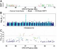
          Chondrodystrophy in dogs is defined by dysplastic, shortened long bones and premature degeneration and calcification of intervertebral discs. Independent genome-wide association analyses for skeletal dysplasia (short limbs) within a single breed ( P Bonferroni = 0.01) and intervertebral disc disease (IVDD) across breeds ( P Bonferroni = 4.0 × 10 −10) both identified a significant association to the same region on CFA12. Whole genome sequencing identified a highly expressed FGF4 retrogene within this shared region. The FGF4 retrogene segregated with limb length and had an odds ratio of 51.23 (95% CI = 46.69, 56.20) for IVDD. Long bone length in dogs is a unique example of multiple disease-causing retrocopies of the same parental gene in a mammalian species. FGF signaling abnormalities have been associated with skeletal dysplasia in humans, and our findings present opportunities for both selective elimination of a medically and financially devastating disease in dogs and further understanding of the ever-growing complexity of retrogene biology.

          Related collections

          Most cited references53

          • Record: found
          • Abstract: found
          • Article: not found

          Active human retrotransposons: variation and disease.

          Mobile DNAs, also known as transposons or 'jumping genes', are widespread in nature and comprise an estimated 45% of the human genome. Transposons are divided into two general classes based on their transposition intermediate (DNA or RNA). Only one subclass, the non-LTR retrotransposons, which includes the Long INterspersed Element-1 (LINE-1 or L1), is currently active in humans as indicated by 96 disease-causing insertions. The autonomous LINE-1 is capable of retrotransposing not only a copy of its own RNA in cis but also other RNAs (Alu, SINE-VNTR-Alu (SVA), U6) in trans to new genomic locations through an element encoded reverse transcriptase. L1 can also retrotranspose cellular mRNAs, resulting in processed pseudogene formation. Here, we highlight recent reports that update our understanding of human L1 retrotransposition and their role in disease. Finally we discuss studies that provide insights into the past and current activity of these retrotransposons, and shed light on not just when, but where, retrotransposition occurs and its part in genetic variation. Copyright © 2012 Elsevier Ltd. All rights reserved.
            Bookmark
            • Record: found
            • Abstract: found
            • Article: found
            Is Open Access

            Nosology and Classification of Genetic Skeletal Disorders: 2010 Revision

            Genetic disorders involving the skeletal system arise through disturbances in the complex processes of skeletal development, growth and homeostasis and remain a diagnostic challenge because of their variety. The Nosology and Classification of Genetic Skeletal Disorders provides an overview of recognized diagnostic entities and groups them by clinical and radiographic features and molecular pathogenesis. The aim is to provide the Genetics, Pediatrics and Radiology community with a list of recognized genetic skeletal disorders that can be of help in the diagnosis of individual cases, in the delineation of novel disorders, and in building bridges between clinicians and scientists interested in skeletal biology. In the 2010 revision, 456 conditions were included and placed in 40 groups defined by molecular, biochemical, and/or radiographic criteria. Of these conditions, 316 were associated with mutations in one or more of 226 different genes, ranging from common, recurrent mutations to “private” found in single families or individuals. Thus, the Nosology is a hybrid between a list of clinically defined disorders, waiting for molecular clarification, and an annotated database documenting the phenotypic spectrum produced by mutations in a given gene. The Nosology should be useful for the diagnosis of patients with genetic skeletal diseases, particularly in view of the information flood expected with the novel sequencing technologies; in the delineation of clinical entities and novel disorders, by providing an overview of established nosologic entities; and for scientists looking for the clinical correlates of genes, proteins and pathways involved in skeletal biology. © 2011 Wiley-Liss, Inc.
              Bookmark
              • Record: found
              • Abstract: found
              • Article: not found

              Mechanisms underlying differential responses to FGF signaling.

              Fibroblast growth factors (FGFs) are key regulators of several developmental processes in which cell fate and differentiation to various tissue lineages are determined. The importance of the proper spatial and temporal regulation of FGF signals is evident from human and mouse genetic studies which show that mutations leading to the dysregulation of FGF signals cause a variety of developmental disorders including dominant skeletal diseases and cancer. The FGF ligands signal via a family of receptor tyrosine kinases and, depending on the cell type or stage of maturation, produce diverse biological responses that include proliferation, growth arrest, differentiation or apoptosis. A central issue in FGF biology is to understand how these diverse cellular responses are determined and how similar signaling inputs can generate distinct patterns of gene expression that govern the specificity of the cellular response. In this review we draw upon studies from the past fifteen years and attempt to construct a molecular picture of the different levels of regulation by which such specific cellular responses could be achieved by FGF signals. We discuss whether specificity could lie in the nature of the ligand, the particular receptor, the signal transduction pathways utilized, or the transcriptional regulation of specific genes. Finally, we also discuss how the interplay of FGF signals with other signaling systems could contribute to the cellular response. In particular we focus on the interaction with the Wnt pathway since FGF/Wnt cross-talk is emerging as an important nexus in regulating a variety of biological processes.
                Bookmark

                Author and article information

                Journal
                Proc Natl Acad Sci U S A
                Proc. Natl. Acad. Sci. U.S.A
                pnas
                pnas
                PNAS
                Proceedings of the National Academy of Sciences of the United States of America
                National Academy of Sciences
                0027-8424
                1091-6490
                24 October 2017
                11 October 2017
                11 October 2017
                : 114
                : 43
                : 11476-11481
                Affiliations
                [1] aDepartment of Population Health and Reproduction, School of Veterinary Medicine, University of California, Davis , CA 95616;
                [2] bDepartment of Surgical and Radiological Sciences, School of Veterinary Medicine, University of California, Davis , CA 95616;
                [3] cDepartment of Animal Science, University of California, Davis , CA 95616;
                [4] dGenome Center, University of California, Davis , CA 95616;
                [5] eBarney and Russum Animal Clinic , Fairfield, CA 94533;
                [6] fDepartment of Veterinary Integrative Biosciences, Texas A&M University , College Station, TX 77843
                Author notes
                1To whom correspondence should be addressed. Email: dlbannasch@ 123456ucdavis.edu .

                Edited by Harris A. Lewin, University of California, Davis, CA, and approved September 18, 2017 (received for review June 2, 2017)

                Author contributions: E.A.B. and D.L.B. designed research; E.A.B., P.J.D., T.M., B.K.S., M.A., A.E.Y., C.K., J.L., C.L.E., and D.L.B. performed research; P.J.D., B.K.S., S.V., R.P., C.T.B., T.R., and D.L.B. contributed new reagents/analytic tools; E.A.B. and D.L.B. analyzed data; and E.A.B., P.J.D., and D.L.B. wrote the paper.

                Author information
                http://orcid.org/0000-0002-4181-4233
                Article
                201709082
                10.1073/pnas.1709082114
                5664524
                29073074
                74373fcc-38ea-4990-83b6-915084e09058
                Copyright © 2017 the Author(s). Published by PNAS.

                This is an open access article distributed under the PNAS license.

                History
                Page count
                Pages: 6
                Funding
                Funded by: HHS | National Institutes of Health (NIH) 100000002
                Award ID: T35 OD010956
                Funded by: HHS | National Institutes of Health (NIH) 100000002
                Award ID: T32 OD010931
                Funded by: UC Davis Signature Research in Genomics
                Award ID: SRG_Bannasch
                Funded by: HHS | NIH | National Institute of Dental and Craniofacial Research (NIDCR) 100000072
                Award ID: R01DE22532
                Funded by: Maxine Adler Endowed Chair Funds
                Award ID: MAC_2016
                Funded by: Center For Companion Animal Health
                Award ID: 2016_20F
                Categories
                Biological Sciences
                Genetics
                From the Cover

                gwas,inherited,genetic,dysplasia,chondrodysplasia
                gwas, inherited, genetic, dysplasia, chondrodysplasia

                Comments

                Comment on this article