2
views
0
recommends
+1 Recommend
0 collections
    0
    shares
      • Record: found
      • Abstract: found
      • Article: found
      Is Open Access

      A role for CBFβ in maintaining the metastatic phenotype of breast cancer cells

      research-article

      Read this article at

      Bookmark
          There is no author summary for this article yet. Authors can add summaries to their articles on ScienceOpen to make them more accessible to a non-specialist audience.

          Abstract

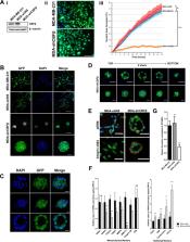
          Epithelial to mesenchymal transition (EMT) is a dynamic process that drives cancer cell plasticity and is thought to play a major role in metastasis. Here we show, using MDA-MB-231 cells as a model, that the plasticity of at least some metastatic breast cancer cells is dependent on the transcriptional co-regulator CBFβ. We demonstrate that CBFβ is essential to maintain the mesenchymal phenotype of triple-negative breast cancer cells and that CBFβ-depleted cells undergo a mesenchymal to epithelial transition (MET) and re-organise into acini-like structures, reminiscent of those formed by epithelial breast cells. We subsequently show, using an inducible CBFβ system, that the MET can be reversed, thus demonstrating the plasticity of CBFβ-mediated EMT. Moreover, the MET can be reversed by expression of the EMT transcription factor Slug whose expression is dependent on CBFβ. Finally, we demonstrate that loss of CBFβ inhibits the ability of metastatic breast cancer cells to invade bone cell cultures and suppresses their ability to form bone metastases in vivo. Together our findings demonstrate that CBFβ can determine the plasticity of the metastatic cancer cell phenotype, suggesting that its regulation in different micro-environments may play a key role in the establishment of metastatic tumours.

          Related collections

          Most cited references21

          • Record: found
          • Abstract: found
          • Article: not found

          Morphogenesis and oncogenesis of MCF-10A mammary epithelial acini grown in three-dimensional basement membrane cultures.

          The three-dimensional culture of MCF-10A mammary epithelial cells on a reconstituted basement membrane results in formation of polarized, growth-arrested acini-like spheroids that recapitulate several aspects of glandular architecture in vivo. Oncogenes introduced into MCF-10A cells disrupt this morphogenetic process, and elicit distinct morphological phenotypes. Recent studies analyzing the mechanistic basis for phenotypic heterogeneity observed among different oncogenes (e.g., ErbB2, cyclin D1) have illustrated the utility of this three-dimensional culture system in modeling the biological activities of cancer genes, particularly with regard to their ability to disrupt epithelial architecture during the early aspects of carcinoma formation. Here we provide a collection of protocols to culture MCF-10A cells, to establish stable pools expressing a gene of interest via retroviral infection, as well as to grow and analyze MCF-10A cells in three-dimensional basement membrane culture.
            Bookmark
            • Record: found
            • Abstract: found
            • Article: not found

            Regulation of breast cancer stem cell activity by signaling through the Notch4 receptor.

            Notch receptor signaling pathways play an important role not only in normal breast development but also in breast cancer development and progression. We assessed the role of Notch receptors in stem cell activity in breast cancer cell lines and nine primary human tumor samples. Stem cells were enriched by selection of anoikis-resistant cells or cells expressing the membrane phenotype ESA(+)/CD44(+)/CD24(low). Using these breast cancer stem cell populations, we compared the activation status of Notch receptors with the status in luminally differentiated cells, and we evaluated the consequences of pathway inhibition in vitro and in vivo. We found that Notch4 signaling activity was 8-fold higher in stem cell-enriched cell populations compared with differentiated cells, whereas Notch1 signaling activity was 4-fold lower in the stem cell-enriched cell populations. Pharmacologic or genetic inhibition of Notch1 or Notch4 reduced stem cell activity in vitro and reduced tumor formation in vivo, but Notch4 inhibition produced a more robust effect with a complete inhibition of tumor initiation observed. Our findings suggest that Notch4-targeted therapies will be more effective than targeting Notch1 in suppressing breast cancer recurrence, as it is initiated by breast cancer stem cells.
              Bookmark
              • Record: found
              • Abstract: found
              • Article: not found

              The RUNX family: developmental regulators in cancer.

              RUNX proteins belong to a family of metazoan transcription factors that serve as master regulators of development. They are frequently deregulated in human cancers, indicating a prominent and, at times, paradoxical role in cancer pathogenesis. The contextual cues that direct RUNX function represent a fast-growing field in cancer research and could provide insights that are applicable to early cancer detection and treatment. This Review describes how RUNX proteins communicate with key signalling pathways during the multistep progression to malignancy; in particular, we highlight the emerging partnership of RUNX with p53 in cancer suppression.
                Bookmark

                Author and article information

                Contributors
                i.holen@sheffield.ac.uk
                paul.shore@manchester.ac.uk
                Journal
                Oncogene
                Oncogene
                Oncogene
                Nature Publishing Group UK (London )
                0950-9232
                1476-5594
                31 January 2020
                31 January 2020
                2020
                : 39
                : 12
                : 2624-2637
                Affiliations
                [1 ]ISNI 0000000121662407, GRID grid.5379.8, Faculty of Biology, Medicine and Health, , University of Manchester, ; Michael Smith Building, Oxford Road, Manchester, M13 9PT UK
                [2 ]ISNI 0000 0000 9868 173X, GRID grid.412787.f, Wuhan University of Science and Technology, ; Jishi Rd, Hongshan Qu, Wuhan Shi, Hubei Sheng, 430065 China
                [3 ]ISNI 0000 0001 2097 4281, GRID grid.29857.31, Penn State University, ; 428 South Frear Laboratory, University Park, Philadelphia, PA 16802 USA
                [4 ]ISNI 0000 0004 1936 9262, GRID grid.11835.3e, Department of Oncology and Metabolism, , University of Sheffield, ; Beech Hill Road, Sheffield, S10 2RX UK
                [5 ]ISNI 0000 0000 8821 5196, GRID grid.23636.32, CRUK Beatson Institute, ; Garscube Estate, Bearsden, Glasgow, G61 1BD UK
                [6 ]ISNI 0000 0001 2193 314X, GRID grid.8756.c, Institute of Cancer Sciences, , University of Glasgow, ; Glasgow, G61 1QH UK
                Author information
                http://orcid.org/0000-0002-4183-8147
                http://orcid.org/0000-0002-4826-0771
                Article
                1170
                10.1038/s41388-020-1170-2
                7082223
                32005976
                73c6ce8f-6485-40ca-8715-07435d137a3d
                © The Author(s) 2020

                Open Access This article is licensed under a Creative Commons Attribution 4.0 International License, which permits use, sharing, adaptation, distribution and reproduction in any medium or format, as long as you give appropriate credit to the original author(s) and the source, provide a link to the Creative Commons license, and indicate if changes were made. The images or other third party material in this article are included in the article’s Creative Commons license, unless indicated otherwise in a credit line to the material. If material is not included in the article’s Creative Commons license and your intended use is not permitted by statutory regulation or exceeds the permitted use, you will need to obtain permission directly from the copyright holder. To view a copy of this license, visit http://creativecommons.org/licenses/by/4.0/.

                History
                : 6 June 2019
                : 4 December 2019
                : 20 January 2020
                Funding
                Funded by: FundRef https://doi.org/10.13039/100009794, Breast Cancer Now (BCN);
                Award ID: May2013
                Award ID: May2013
                Award ID: May2013
                Award Recipient :
                Funded by: FundRef https://doi.org/10.13039/501100000289, Cancer Research UK (CRUK);
                Award ID: PhD Studentship
                Award Recipient :
                Categories
                Article
                Custom metadata
                © Springer Nature Limited 2020

                Oncology & Radiotherapy
                cancer,mechanisms of disease,breast cancer
                Oncology & Radiotherapy
                cancer, mechanisms of disease, breast cancer

                Comments

                Comment on this article