5
views
0
recommends
+1 Recommend
1 collections
    0
    shares
      • Record: found
      • Abstract: found
      • Article: found

      Dihydropyridine receptor (DHPR, CACNA1S) congenital myopathy

      , , , , , , , , , , , , , , , , , , , , , , , , , , , , , , , , , , , , , , ,
      Acta Neuropathologica
      Springer Science and Business Media LLC

      Read this article at

      ScienceOpenPublisherPubMed
      Bookmark
          There is no author summary for this article yet. Authors can add summaries to their articles on ScienceOpen to make them more accessible to a non-specialist audience.

          Abstract

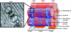
          Muscle contraction upon nerve stimulation relies on excitation-contraction coupling (ECC) to promote the rapid and generalized release of calcium within myofibers. In skeletal muscle, ECC is performed by the direct coupling of a voltage-gated L-type Ca(2+) channel (dihydropyridine receptor; DHPR) located on the T-tubule with a Ca(2+) release channel (ryanodine receptor; RYR1) on the sarcoplasmic reticulum (SR) component of the triad. Here, we characterize a novel class of congenital myopathy at the morphological, molecular, and functional levels. We describe a cohort of 11 patients from 7 families presenting with perinatal hypotonia, severe axial and generalized weakness. Ophthalmoplegia is present in four patients. The analysis of muscle biopsies demonstrated a characteristic intermyofibrillar network due to SR dilatation, internal nuclei, and areas of myofibrillar disorganization in some samples. Exome sequencing revealed ten recessive or dominant mutations in CACNA1S (Cav1.1), the pore-forming subunit of DHPR in skeletal muscle. Both recessive and dominant mutations correlated with a consistent phenotype, a decrease in protein level, and with a major impairment of Ca(2+) release induced by depolarization in cultured myotubes. While dominant CACNA1S mutations were previously linked to malignant hyperthermia susceptibility or hypokalemic periodic paralysis, our findings strengthen the importance of DHPR for perinatal muscle function in human. These data also highlight CACNA1S and ECC as therapeutic targets for the development of treatments that may be facilitated by the previous knowledge accumulated on DHPR.

          Related collections

          Most cited references34

          • Record: found
          • Abstract: found
          • Article: not found

          Structure of the voltage-gated calcium channel Cav1.1 complex.

          The voltage-gated calcium channel Ca(v)1.1 is engaged in the excitation-contraction coupling of skeletal muscles. The Ca(v)1.1 complex consists of the pore-forming subunit α1 and auxiliary subunits α2δ, β, and γ. We report the structure of the rabbit Ca(v)1.1 complex determined by single-particle cryo-electron microscopy. The four homologous repeats of the α1 subunit are arranged clockwise in the extracellular view. The γ subunit, whose structure resembles claudins, interacts with the voltage-sensing domain of repeat IV (VSD(IV)), whereas the cytosolic β subunit is located adjacent to VSD(II) of α1. The α2 subunit interacts with the extracellular loops of repeats I to III through its VWA and Cache1 domains. The structure reveals the architecture of a prototypical eukaryotic Ca(v) channel and provides a framework for understanding the function and disease mechanisms of Ca(v) and Na(v) channels.
            Bookmark
            • Record: found
            • Abstract: found
            • Article: found
            Is Open Access

            T-tubule biogenesis and triad formation in skeletal muscle and implication in human diseases

            In skeletal muscle, the excitation-contraction (EC) coupling machinery mediates the translation of the action potential transmitted by the nerve into intracellular calcium release and muscle contraction. EC coupling requires a highly specialized membranous structure, the triad, composed of a central T-tubule surrounded by two terminal cisternae from the sarcoplasmic reticulum. While several proteins located on these structures have been identified, mechanisms governing T-tubule biogenesis and triad formation remain largely unknown. Here, we provide a description of triad structure and plasticity and review the role of proteins that have been linked to T-tubule biogenesis and triad formation and/or maintenance specifically in skeletal muscle: caveolin 3, amphiphysin 2, dysferlin, mitsugumins, junctophilins, myotubularin, ryanodine receptor, and dihydhropyridine Receptor. The importance of these proteins in triad biogenesis and subsequently in muscle contraction is sustained by studies on animal models and by the direct implication of most of these proteins in human myopathies.
              Bookmark
              • Record: found
              • Abstract: found
              • Article: not found

              Muscle weakness in myotonic dystrophy associated with misregulated splicing and altered gating of Ca(V)1.1 calcium channel.

              Myotonic dystrophy type 1 and type 2 (DM1 and DM2) are genetic diseases in which mutant transcripts containing expanded CUG or CCUG repeats cause cellular dysfunction by altering the processing or metabolism of specific mRNAs and miRNAs. The toxic effects of mutant RNA are mediated partly through effects on proteins that regulate alternative splicing. Here we show that alternative splicing of exon 29 (E29) of Ca(V)1.1, a calcium channel that controls skeletal muscle excitation-contraction coupling, is markedly repressed in DM1 and DM2. The extent of E29 skipping correlated with severity of weakness in tibialis anterior muscle of DM1 patients. Two splicing factors previously implicated in DM1, MBNL1 and CUGBP1, participated in the regulation of E29 splicing. In muscle fibers of wild-type mice, the Ca(V)1.1 channel conductance and voltage sensitivity were increased by splice-shifting oligonucleotides that induce E29 skipping. In contrast to human DM1, expression of CUG-expanded RNA caused only a modest increase in E29 skipping in mice. However, forced skipping of E29 in these mice, to levels approaching those observed in human DM1, aggravated the muscle pathology as evidenced by increased central nucleation. Together, these results indicate that DM-associated splicing defects alter Ca(V)1.1 function, with potential for exacerbation of myopathy.
                Bookmark

                Author and article information

                Journal
                Acta Neuropathologica
                Acta Neuropathol
                Springer Science and Business Media LLC
                0001-6322
                1432-0533
                April 2017
                December 23 2016
                April 2017
                : 133
                : 4
                : 517-533
                Article
                10.1007/s00401-016-1656-8
                28012042
                71e44b05-7df3-4da0-910a-bb89c55d50a1
                © 2017

                http://www.springer.com/tdm

                History

                Comments

                Comment on this article