1
views
0
recommends
+1 Recommend
0 collections
    0
    shares
      • Record: found
      • Abstract: found
      • Article: found
      Is Open Access

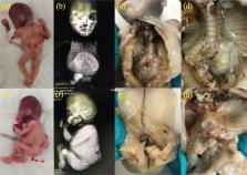
      Prenatally diagnosed fetal thoraco-lumbar spine duplication associated with lipomyelomeningocele: An extremely rare case of split cord malformation

      case-report

      Read this article at

      Bookmark
          There is no author summary for this article yet. Authors can add summaries to their articles on ScienceOpen to make them more accessible to a non-specialist audience.

          Abstract

          Spine duplication is considered rare, a more serious form of split cord malformation. Ultrasonographic evaluation of the spine in the second trimester is central to the antenatal diagnosis of spinal malformations. Here, we report a case of thoraco-lumbar spine duplication associated with lipomyelomeningocele diagnosed by ultrasonography at 19 weeks of gestation. To the best of our knowledge, this is the first case report of spine duplication diagnosed by antenatal ultrasonography.

          Related collections

          Most cited references18

          • Record: found
          • Abstract: found
          • Article: not found

          Split cord malformation: Part I: A unified theory of embryogenesis for double spinal cord malformations.

          Much confusion still exists concerning the pathological definitions and clinical significance of double spinal cord malformations. Traditional terms used to describe the two main forms of these rare malformations, diastematomyelia and diplomyelia, add to the confusion by their inconsistent usage, ambiguities, and implications of their dissimilar embryogenesis. Based on the detailed radiographic and surgical findings of 39 cases of double cord malformations and the autopsy data on two other cases, this study endorses a new classification for double cord malformations and proposes a unified theory of embryogenesis for all their variant forms and features. The new classification recommends the term split cord malformation (SCM) for all double spinal cords. A Type I SCM consists of two hemicords, each contained within its own dural tube and separated by a dura-sheathed rigid osseocartilaginous median septum. A Type II SCM consists of two hemicords housed in a single dural tube separated by a nonrigid, fibrous median septum. These two essential features necessary for typing, the state of the dural tube and the nature of the median septum, do not ever overlap between the two main forms and can always be demonstrated by imaging studies so that accurate preoperative typing is always possible. All other associated structures in SCM such as paramedian nerve roots, myelomeningoceles manqué, and centromedian vascular structures frequently do overlap between types and are not reliable typing criteria. The unified theory of embryogenesis proposes that all variant types of SCMs have a common embryogenetic mechanism. Basic to this mechanism is the formation of adhesions between ecto- and endoderm, leading to an accessory neurenteric canal around which condenses an endomesenchymal tract that bisects the developing notochord and causes formation of two hemineural plates. The altered state of the emerging split neural tube and the subsequent ontogenetic fates of the constituent components of the endomesenchymal tract ultimately determine the configuration and orientation of the hemicords, the nature of the median septum, the coexistence of various vascular, lipomatous, neural, and fibrous oddities within the median cleft, the high association with open myelodysplastic and cutaneous lesions, and the seemingly unlikely relationship with fore and midgut anomalies. The multiple facets of this theory are presented in increasing complexity against the background of known embryological facts and theories; the validity of each facet is tested by comparing structures and phenomena predicted by the facet with actual radiographic, surgical, and histopathological findings of these 41 cases of SCM.
            Bookmark
            • Record: found
            • Abstract: found
            • Article: not found

            Split cord malformation: Part II: Clinical syndrome.

            Raina Pang (1992)
            Thirty-nine patients with split cord malformations (SCM) were studied in detail with respect to their clinical, radiographic, and surgical findings as well as their outcome data. Eight patients were adults and 31 patients were children. According to the classification endorsed by Part I of the SCM study, 19 patients had Type I SCM (6 adults and 13 children), 18 patients had Type II SCM (2 adults and 16 children), and 2 patients had composite SCM with both lesion types situated in tandem. Six SCMs were cervical, 2 were thoracic, and 31 were in the lumbar region. All 8 adults had pain and progressive sensorimotor deficits at diagnosis. Only 16 of the 31 children had symptoms, and among these, 14 had progressive sensorimotor deficits, but only 6 had pain. The difference in the clinical picture between adults and children is similar to that described in the tethered cord syndrome, except for left-right functional discrepancy, which was prominent in 8 children with SCM but rarely seen in tethered cord syndrome due to other causes. Cutaneous manifestations of either occult or open dysraphic states were present in all but 3 patients; hypertrichosis was by far the best predictor of an underlying SCM, being found in 56% in the series. Neurological deterioration in SCM was independent of the lesion type: the Type I:Type II ratio for symptomatic progression was 13:11. It was also independent of the location of the lesion: 67% of patients with cervical SCMs had symptomatic progression versus 64% of patients with thoracolumbar lesions. High-resolution, thin cut, axial computed tomographic myelography using bone algorithms was more sensitive than magnetic resonance imaging in defining the anatomical details of the SCM. Radiographic classifications of the SCM, using the nature of the median septum and the number of dural tubes as criteria, was always possible without ambiguity. However, whereas every Type I bone septum was identified preoperatively, only 5 Type II fibrous septa were revealed by preoperative imaging, even though a fibrous septum and/or other fibroneurovascular bands were found tethering the hemicords in every Type II case at surgery. Complete imaging studies also showed that all lumbar SCMs had low-lying coni and at least one additional tethering lesion besides the split cords, whereas only 1 of 7 cervical and high thoracic SCMs had a low conus and a second tethering lesion. The surgical goal for SCM was release of the tethered hemicords by eliminating the bone spurs, dural sleeves, fibrous septa, or any fibroneurovascular bands (myelomeningoceles manqué) that might be transfixing the split cord. Type I cases were technically more difficult and had a slightly higher surgical morbidity than Type II cases, especially if an oblique bone septum had asymmetrically divided the cord into one larger hemicord and one smaller, hence, very delicate, hemicord.(ABSTRACT TRUNCATED AT 400 WORDS)
              Bookmark
              • Record: found
              • Abstract: found
              • Article: not found

              Increased DNA methylation at the AXIN1 gene in a monozygotic twin from a pair discordant for a caudal duplication anomaly.

              The AXIN1 gene has been implicated in caudal duplication anomalies. Its coding region was sequenced in both members of a monozygotic (MZ) twin pair discordant for a caudal duplication anomaly, but no mutation was found. Using bisulfite sequencing, we examined methylation at the promoter region of the AXIN1 gene in these twins and in twin and age-matched singleton controls. Methylation of the promoter region in peripheral blood mononucleated cells was variable among individuals, including MZ pairs. In the MZ pair discordant for the caudal duplication, this region of the affected twin was significantly more methylated than that of the unaffected twin (P < .0001), which was significantly more methylated than those of the controls (P = .02). We have confirmed that this CpG island does function as a promoter in vitro and that its activity is inversely proportional to the extent of methylation. This finding raises the possibility that hypermethylation of the AXIN1 promoter, by mechanisms as yet undetermined, is associated with the malformation. This case may be paradigmatic for some cases of MZ discordance.
                Bookmark

                Author and article information

                Journal
                Turk J Obstet Gynecol
                Turk J Obstet Gynecol
                TJOG
                Turkish Journal of Obstetrics and Gynecology
                Galenos Publishing
                2149-9322
                2149-9330
                December 2022
                13 December 2022
                : 19
                : 4
                : 333-337
                Affiliations
                [1 ]Marmara University Pendik Training and Research Hospital, Clinic of Perinatology, İstanbul, Turkey
                [2 ]University of Health Sciences Turkey, Zeynep Kamil Women’s and Children’s Disease Training and Research Hospital, Clinic of Perinatology, İstanbul, Turkey
                [3 ]University of Health Sciences Turkey, Zeynep Kamil Women’s and Children’s Disease Training and Research Hospital, Clinic of Pathology, İstanbul, Turkey
                Author notes
                * Address for Correspondence: Marmara University Pendik Training and Research Hospital, Clinic of Perinatology, İstanbul, Turkey Phone: +90 530 408 48 00 E-mail: munipakalin89@ 123456gmail.com
                Author information
                https://orcid.org/0000-0002-3737-7712
                https://orcid.org/0000-0001-5578-4437
                https://orcid.org/0000-0003-2002-0386
                https://orcid.org/0000-0001-7255-860X
                Article
                57530
                10.4274/tjod.galenos.2022.85453
                9748855
                36511649
                7197ef1b-5863-499d-96a8-54695b6a2945
                ©Copyright 2022 by Turkish Society of Obstetrics and Gynecology | Turkish Journal of Obstetrics and Gynecology published by Galenos Publishing House.

                This is an open-access article distributed under the terms of the Creative Commons Attribution License, which permits unrestricted use, distribution, and reproduction in any medium, provided the original work is properly cited.

                History
                : 7 August 2022
                : 16 October 2022
                Categories
                Case Report

                congenital spine defect,spinal dysraphism,spine duplication,split cord malformations,split notochord syndrome

                Comments

                Comment on this article