6
views
0
recommends
+1 Recommend
0 collections
    0
    shares
      • Record: found
      • Abstract: found
      • Article: found
      Is Open Access

      EEG microstate features according to performance on a mental arithmetic task

      research-article
      , , ,
      Scientific Reports
      Nature Publishing Group UK
      Cognitive neuroscience, Imaging

      Read this article at

      Bookmark
          There is no author summary for this article yet. Authors can add summaries to their articles on ScienceOpen to make them more accessible to a non-specialist audience.

          Abstract

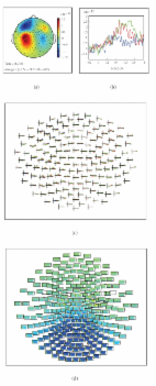
          In this study, we hypothesized that task performance could be evaluated applying EEG microstate to mental arithmetic task. This pilot study also aimed at evaluating the efficacy of microstates as novel features to discriminate task performance. Thirty-six subjects were divided into good and poor performers, depending on how well they performed the task. Microstate features were derived from EEG recordings during resting and task states. In the good performers, there was a decrease in type C and an increase in type D features during the task compared to the resting state. Mean duration and occurrence decreased and increased, respectively. In the poor performers, occurrence of type D feature, mean duration and occurrence showed greater changes. We investigated whether microstate features were suitable for task performance classification and eleven features including four archetypes were selected by recursive feature elimination (RFE). The model that implemented them showed the highest classification performance for differentiating between groups. Our pilot findings showed that the highest mean Area Under Curve (AUC) was 0.831. This study is the first to apply EEG microstate features to specific cognitive tasks in healthy subjects, suggesting that EEG microstate features can reflect task achievement.

          Related collections

          Most cited references78

          • Record: found
          • Abstract: found
          • Article: found
          Is Open Access

          FieldTrip: Open Source Software for Advanced Analysis of MEG, EEG, and Invasive Electrophysiological Data

          This paper describes FieldTrip, an open source software package that we developed for the analysis of MEG, EEG, and other electrophysiological data. The software is implemented as a MATLAB toolbox and includes a complete set of consistent and user-friendly high-level functions that allow experimental neuroscientists to analyze experimental data. It includes algorithms for simple and advanced analysis, such as time-frequency analysis using multitapers, source reconstruction using dipoles, distributed sources and beamformers, connectivity analysis, and nonparametric statistical permutation tests at the channel and source level. The implementation as toolbox allows the user to perform elaborate and structured analyses of large data sets using the MATLAB command line and batch scripting. Furthermore, users and developers can easily extend the functionality and implement new algorithms. The modular design facilitates the reuse in other software packages.
            Bookmark
            • Record: found
            • Abstract: found
            • Article: not found

            A default mode of brain function.

            A baseline or control state is fundamental to the understanding of most complex systems. Defining a baseline state in the human brain, arguably our most complex system, poses a particular challenge. Many suspect that left unconstrained, its activity will vary unpredictably. Despite this prediction we identify a baseline state of the normal adult human brain in terms of the brain oxygen extraction fraction or OEF. The OEF is defined as the ratio of oxygen used by the brain to oxygen delivered by flowing blood and is remarkably uniform in the awake but resting state (e.g., lying quietly with eyes closed). Local deviations in the OEF represent the physiological basis of signals of changes in neuronal activity obtained with functional MRI during a wide variety of human behaviors. We used quantitative metabolic and circulatory measurements from positron-emission tomography to obtain the OEF regionally throughout the brain. Areas of activation were conspicuous by their absence. All significant deviations from the mean hemisphere OEF were increases, signifying deactivations, and resided almost exclusively in the visual system. Defining the baseline state of an area in this manner attaches meaning to a group of areas that consistently exhibit decreases from this baseline, during a wide variety of goal-directed behaviors monitored with positron-emission tomography and functional MRI. These decreases suggest the existence of an organized, baseline default mode of brain function that is suspended during specific goal-directed behaviors.
              Bookmark
              • Record: found
              • Abstract: found
              • Article: not found

              PhysioBank, PhysioToolkit, and PhysioNet

              Circulation, 101(23)
                Bookmark

                Author and article information

                Contributors
                leebr@gist.ac.kr
                Journal
                Sci Rep
                Sci Rep
                Scientific Reports
                Nature Publishing Group UK (London )
                2045-2322
                11 January 2021
                11 January 2021
                2021
                : 11
                : 343
                Affiliations
                GRID grid.61221.36, ISNI 0000 0001 1033 9831, Department of Biomedical Science and Engineering (BMSE), Institute Integrated Technology (IIT), , Gwangju Institute of Science and Technology (GIST), ; 123 Cheomdan-gwagiro, Buk-gu, Gwangju, 61005 South Korea
                Article
                79423
                10.1038/s41598-020-79423-7
                7801706
                33431963
                7161dc8f-a9a8-4903-a7b2-604b97968b61
                © The Author(s) 2021

                Open Access This article is licensed under a Creative Commons Attribution 4.0 International License, which permits use, sharing, adaptation, distribution and reproduction in any medium or format, as long as you give appropriate credit to the original author(s) and the source, provide a link to the Creative Commons licence, and indicate if changes were made. The images or other third party material in this article are included in the article's Creative Commons licence, unless indicated otherwise in a credit line to the material. If material is not included in the article's Creative Commons licence and your intended use is not permitted by statutory regulation or exceeds the permitted use, you will need to obtain permission directly from the copyright holder. To view a copy of this licence, visit http://creativecommons.org/licenses/by/4.0/.

                History
                : 5 June 2020
                : 30 November 2020
                Funding
                Funded by: FundRef http://dx.doi.org/10.13039/501100003725, National Research Foundation of Korea;
                Award ID: 2020R1A2B5B01002297
                Categories
                Article
                Custom metadata
                © The Author(s) 2021

                Uncategorized
                cognitive neuroscience,imaging
                Uncategorized
                cognitive neuroscience, imaging

                Comments

                Comment on this article