272
views
0
recommends
+1 Recommend
0 collections
    0
    shares
      • Record: found
      • Abstract: found
      • Article: found
      Is Open Access

      Dynamic Regulation of Sarcomeric Actin Filaments in Striated Muscle

      review-article

      Read this article at

      Bookmark
          There is no author summary for this article yet. Authors can add summaries to their articles on ScienceOpen to make them more accessible to a non-specialist audience.

          Abstract

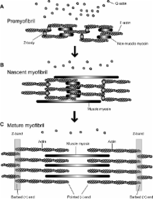
          In striated muscle, the actin cytoskeleton is differentiated into myofibrils. Actin and myosin filaments are organized in sarcomeres and specialized for producing contractile forces. Regular arrangement of actin filaments with uniform length and polarity is critical for the contractile function. However, the mechanisms of assembly and maintenance of sarcomeric actin filaments in striated muscle are not completely understood. Live imaging of actin in striated muscle has revealed that actin subunits within sarcomeric actin filaments are dynamically exchanged without altering overall sarcomeric structures. A number of regulators for actin dynamics have been identified, and malfunction of these regulators often result in disorganization of myofibril structures or muscle diseases. Therefore, proper regulation of actin dynamics in striated muscle is critical for assembly and maintenance of functional myofibrils. Recent studies have suggested that both enhancers of actin dynamics and stabilizers of actin filaments are important for sarcomeric actin organization. Further investigation of the regulatory mechanism of actin dynamics in striated muscle should be a key to understanding how myofibrils develop and operate. © 2010 Wiley-Liss, Inc.

          Related collections

          Most cited references254

          • Record: found
          • Abstract: found
          • Article: not found

          Actin, a central player in cell shape and movement.

          The protein actin forms filaments that provide cells with mechanical support and driving forces for movement. Actin contributes to biological processes such as sensing environmental forces, internalizing membrane vesicles, moving over surfaces, and dividing the cell in two. These cellular activities are complex; they depend on interactions of actin monomers and filaments with numerous other proteins. Here, we present a summary of the key questions in the field and suggest how those questions might be answered. Understanding actin-based biological phenomena will depend on identifying the participating molecules and defining their molecular mechanisms. Comparisons of quantitative measurements of reactions in live cells with computer simulations of mathematical models will also help generate meaningful insights.
            Bookmark
            • Record: found
            • Abstract: found
            • Article: not found

            Unleashing formins to remodel the actin and microtubule cytoskeletons.

            Formins are highly conserved proteins that have essential roles in remodelling the actin and microtubule cytoskeletons to influence eukaryotic cell shape and behaviour. Recent work has identified numerous cellular factors that locally recruit, activate or inactivate formins to bridle and unleash their potent effects on actin nucleation and elongation. The effects of formins on microtubules have also begun to be described, which places formins in a prime position to coordinate actin and microtubule dynamics. The emerging complexity in the mechanisms governing formins mirrors the wide range of essential functions that they perform in cell motility, cell division and cell and tissue morphogenesis.
              Bookmark
              • Record: found
              • Abstract: found
              • Article: not found

              Myosin II functions in actin-bundle turnover in neuronal growth cones.

              Retrograde actin flow works in concert with cell adhesion to generate traction forces that are involved in axon guidance in neuronal growth cones. Myosins have been implicated in retrograde flow, but identification of the specific myosin subtype(s) involved has been controversial. Using fluorescent speckle microscopy (FSM) to assess actin dynamics, we report that inhibition of myosin II alone decreases retrograde flow by 51% and the remaining flow can be almost fully accounted for by the 'push' of plus-end actin assembly at the leading edge of the growth cone. Interestingly, actin bundles that are associated with filopodium roots elongated by approximately 83% after inhibition of myosin II. This unexpected result was due to decreased rates of actin-bundle severing near their proximal (minus or pointed) ends which are located in the transition zone of the growth cone. Our study reveals a mechanism for the regulation of actin-bundle length by myosin II that is dependent on actin-bundle severing, and demonstrate that retrograde flow is a steady state that depends on both myosin II contractility and actin-network treadmilling.
                Bookmark

                Author and article information

                Journal
                Cytoskeleton (Hoboken)
                cm
                Cytoskeleton (Hoboken, N.j.)
                John Wiley & Sons, Inc.
                1949-3584
                1949-3592
                November 2010
                09 August 2010
                : 67
                : 11
                : 677-692
                Affiliations
                simpleDepartment of Pathology and Department of Cell Biology, Emory University Atlanta, Georgia
                Author notes
                *Address correspondence to: Shoichiro Ono, Department of Pathology, Emory University, 615 Michael Street, Whitehead Research Building, Room 105N, Atlanta, GA 30322, USA. E-mail: sono@ 123456emory.edu

                Monitoring Editor: Pekka Lappalainen

                Article
                10.1002/cm.20476
                2963174
                20737540
                70e55128-2c94-4060-9100-e36c30f45e3c
                Copyright © 2010 Wiley-Liss, Inc., A Wiley Company

                Re-use of this article is permitted in accordance with the Creative Commons Deed, Attribution 2.5, which does not permit commercial exploitation.

                History
                : 03 June 2010
                : 21 July 2010
                : 29 July 2010
                Categories
                Review Article

                Cell biology
                actin turnover,stabilization,congenital myopathy,depolymerization,myofibrils,sarcomeres,capping

                Comments

                Comment on this article