8
views
0
recommends
+1 Recommend
0 collections
    0
    shares
      • Record: found
      • Abstract: found
      • Article: found
      Is Open Access

      Imaging with ultrasound in physical therapy: What is the PT’s scope of practice? A competency-based educational model and training recommendations

      research-article

      Read this article at

      Bookmark
          There is no author summary for this article yet. Authors can add summaries to their articles on ScienceOpen to make them more accessible to a non-specialist audience.

          Abstract

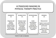
          Physical therapists employ ultrasound (US) imaging technology for a broad range of clinical and research purposes. Despite this, few physical therapy regulatory bodies guide the use of US imaging, and there are limited continuing education opportunities for physical therapists to become proficient in using US within their professional scope of practice. Here, we (i) outline the current status of US use by physical therapists; (ii) define and describe four broad categories of physical therapy US applications (ie, rehabilitation, diagnostic, intervention and research US); (iii) discuss how US use relates to the scope of high value physical therapy practice and (iv) propose a broad framework for a competency-based education model for training physical therapists in US. This paper only discusses US imaging—not ‘therapeutic’ US. Thus, ‘imaging’ is implicit anywhere the term ‘ultrasound’ is used.

          Related collections

          Most cited references61

          • Record: found
          • Abstract: found
          • Article: not found

          Rapid atrophy of the lumbar multifidus follows experimental disc or nerve root injury.

          Experimental study of muscle changes after lumbar spinal injury. To investigate effects of intervertebral disc and nerve root lesions on cross-sectional area, histology and chemistry of porcine lumbar multifidus. The multifidus cross-sectional area is reduced in acute and chronic low back pain. Although chronic changes are widespread, acute changes at 1 segment are identified within days of injury. It is uncertain whether changes precede or follow injury, or what is the mechanism. The multifidus cross-sectional area was measured in 21 pigs from L1 to S1 with ultrasound before and 3 or 6 days after lesions: incision into L3-L4 disc, medial branch transection of the L3 dorsal ramus, and a sham procedure. Samples from L3 to L5 were studied histologically and chemically. The multifidus cross-sectional area was reduced at L4 ipsilateral to disc lesion but at L4-L6 after nerve lesion. There was no change after sham or on the opposite side. Water and lactate were reduced bilaterally after disc lesion and ipsilateral to nerve lesion. Histology revealed enlargement of adipocytes and clustering of myofibers at multiple levels after disc and nerve lesions. These data resolve the controversy that the multifidus cross-sectional area reduces rapidly after lumbar injury. Changes after disc lesion affect 1 level with a different distribution to denervation. Such changes may be due to disuse following reflex inhibitory mechanisms.
            Bookmark
            • Record: found
            • Abstract: found
            • Article: not found

            Effect of stabilization training on multifidus muscle cross-sectional area among young elite cricketers with low back pain.

            A single-blinded, pretreatment-posttreatment assessment. To investigate, using ultrasound imaging, the cross-sectional area (CSA) of the lumbar multifidus muscle at 4 vertebral levels (L2, L3, L4, L5) in elite cricketers with and without low back pain (LBP) and (2) to document the effect of a staged stabilization training program on multifidus muscle CSA. Despite high fitness levels and often intensive strength training programs, athletes still suffer LBP. The incidence of LBP among Australian cricketers is 8% and as high as 14% among fast bowlers. Previous researchers have found that the multifidus muscle contributes to segmental stability of the lumbopelvic region; however, the CSA of this muscle has not been previously assessed in elite cricketers. CSAs of the multifidus muscles were assessed at rest on the left and right sides for 4 vertebral levels at the start and completion of a 13-week cricket training camp. Participants who reported current or previous LBP were placed in a rehabilitation group. The stabilization program involved voluntary contraction of the multifidus, transversus abdominis, and pelvic floor muscles, with real-time feedback from rehabilitative ultrasound imaging (RUSI), progressed from non-weight-bearing to weight-bearing positions and movement training. Pain scores (using a visual analogue scale) were also collected from those with LBP. The CSAs of the multifidus muscles at the L5 vertebral level increased for the 7 cricketers with LBP who received the stabilization training, compared with the 14 cricketers without LBP who did not receive rehabilitation (P = .004). In addition, the amount of muscle asymmetry among those with LBP significantly decreased (P = .029) and became comparable to cricketers without LBP. These effects were not evident for the L2, L3, and L4 vertebral levels. There was also a 50% decrease in the mean reported pain level among the cricketers with LBP. Multifidus muscle atrophy can exist in highly active, elite athletes with LBP. Specific retraining resulted in an improvement in multifidus muscle CSA and this was concomitant with a decrease in pain. Therapy, level 2b.
              Bookmark
              • Record: found
              • Abstract: found
              • Article: not found

              Rehabilitative ultrasound imaging of the abdominal muscles.

              Rehabilitative ultrasound imaging (RUSI) of the abdominal muscles is increasingly being used in the management of conditions involving musculoskeletal dysfunctions associated with the abdominal muscles, including certain types of low back and pelvic pain. This commentary provides an overview of current concepts and evidence related to RUSI of the abdominal musculature, including issues addressing the potential role of ultrasound imaging in the assessment and training of these muscles. Both quantitative and qualitative aspects associated with clinical and research applications are considered, as are the possible limitations related to the interpretation of measurements made with RUSI. Research to date has utilized a range of methodological approaches, including different transducer placements and imaging techniques. The pros and cons of the various methods are discussed, and guidelines for future investigations are presented. Potential implications and opportunities for clinical use of RUSI to enhance evidence-based practice are outlined, as are suggestions for future research to further clarify the possible role of RUSI in the evaluation and treatment of abdominal muscular morphology and function.
                Bookmark

                Author and article information

                Journal
                Br J Sports Med
                Br J Sports Med
                bjsports
                bjsm
                British Journal of Sports Medicine
                BMJ Publishing Group (BMA House, Tavistock Square, London, WC1H 9JR )
                0306-3674
                1473-0480
                December 2019
                25 April 2019
                : 53
                : 23
                : 1447-1453
                Affiliations
                [1 ] departmentDepartment of Physical Therapy, Faculty of Rehabilitation Medicine , University of Alberta , Edmonton, Alberta, Canada
                [2 ] departmentHealth and Rehabilitation Research Institute, School of Clinical Sciences , Auckland University of Technology , Auckland, New Zealand
                [3 ] departmentSchool of Health and Rehabilitation Sciences , The University of Queensland , Brisbane, Queensland, Australia
                [4 ] departmentDepartment of Physiotherapy and Performance Science , University College Dublin , Dublin, Ireland
                [5 ] departmentSchool of Allied Health Sciences , Griffith University , Brisbane, Queensland, Australia
                [6 ] departmentDepartmento de Enfermeria y Fisioterapia , Universidad de Alcala de Henares , Madrid, Spain
                [7 ] departmentDepartamento de Fisioterapia , Universidad Francisco de Victoria , Madrid, Spain
                [8 ] Walter Reed Army Institute of Research , Silver Spring, Maryland, USA
                [9 ] departmentSchool of Health Professions and Rehabilitation Sciences , University of Southampton , Southampton, UK
                Author notes
                [Correspondence to ] Dr Jackie L Whittaker, Faculty of Rehabilitation Medicine, University of Alberta, Edmonton, AB T6G 2G4, Canada; jwhittak@ 123456ualberta.ca
                Author information
                http://orcid.org/0000-0002-6591-4976
                http://orcid.org/0000-0002-1206-9107
                Article
                bjsports-2018-100193
                10.1136/bjsports-2018-100193
                6900235
                31023858
                70a24b5c-7f97-4945-9653-7cd1cf8a46da
                © Author(s) (or their employer(s)) 2019. Re-use permitted under CC BY. Published by BMJ.

                This is an open access article distributed in accordance with the Creative Commons Attribution 4.0 Unported (CC BY 4.0) license, which permits others to copy, redistribute, remix, transform and build upon this work for any purpose, provided the original work is properly cited, a link to the licence is given, and indication of whether changes were made. See: https://creativecommons.org/licenses/by/4.0/.

                History
                : 25 March 2019
                Categories
                Consensus Statement
                1506
                1507
                2314
                Custom metadata
                unlocked
                editors-choice

                Sports medicine
                curriculum,education,professional issues,rehabilitation,sonography
                Sports medicine
                curriculum, education, professional issues, rehabilitation, sonography

                Comments

                Comment on this article