36
views
0
recommends
+1 Recommend
0 collections
    0
    shares
      • Record: found
      • Abstract: found
      • Article: found
      Is Open Access

      PTH/SDF-1α cotherapy induces CD90+CD34− stromal cells migration and promotes tissue regeneration in a rat periodontal defect model

      research-article
      1 , 2 , 1 , 2 , a , 1 , 2
      Scientific Reports
      Nature Publishing Group

      Read this article at

      Bookmark
          There is no author summary for this article yet. Authors can add summaries to their articles on ScienceOpen to make them more accessible to a non-specialist audience.

          Abstract

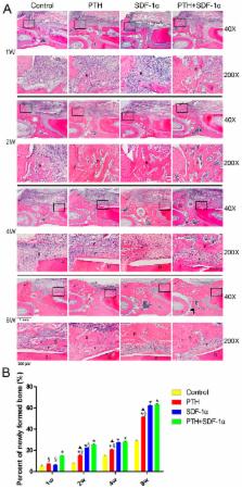
          Stromal cell-derived factor-1α (SDF-1α) is a key stem cell homing factor that is crucial for recruitment of stem cells to many diseased organs. However, the therapeutic activity of SDF-1α is potentially limited by N-terminal cleavage at position-2 proline by a cell surface protein CD26/dipeptidyl peptidase-IV (DPP-IV). Parathyroid hormone (PTH) is a DPP-IV inhibitor and has been suggested as a promising agent for periodontal tissue repair. The purpose of this study was to explore the effects of a cell-free system comprising SDF-1α and scaffold plus PTH systemic application on periodontal tissue regeneration in vivo. The results showed that PTH/SDF-1α cotherapy improved the quantity of regenerated bone and resulted in better organization of ligament interface. We further investigated the possible mechanisms, and found that PTH/SDF-1α cotherapy enhanced CD90+CD34− stromal cells migration in vivo, increased the number of CXCR4 + cells in periodontal defects, induced early bone osteoclastogenesis and enhanced the expression of runt-related transcription factor 2 (Runx2), alkaline phosphatase (ALP) and collagen I (Col I) in newly formed bone tissue. In conclusion, this cell-free tissue engineering system with local administration of SDF-1α and systemic application of PTH could be employed to induce CD90+CD34− stromal cells recruitment and promote periodontal tissue regeneration.

          Related collections

          Most cited references34

          • Record: found
          • Abstract: found
          • Article: not found

          The lymphocyte chemoattractant SDF-1 is a ligand for LESTR/fusin and blocks HIV-1 entry.

          Chemokines are chemotactic cytokines that activate and direct the migration of leukocytes. There are two subfamilies, the CXC and the CC chemokines. We recently found that the CXC-chemokine stromal cell-derived factor-1 (SDF-1) is a highly efficacious lymphocyte chemoattractant. Chemokines act on responsive leukocyte subsets through G-protein-coupled seven-transmembrane receptors, which are also used by distinct strains of HIV-1 as cofactors for viral entry. Laboratory-adapted and some T-cell-line-tropic (T-tropic) primary viruses use the orphan chemokine receptor LESTR/fusin (also known as fusin), whereas macrophage-tropic primary HIV-1 isolates use CCR-5 and CCR-3 (refs 7-11), which are receptors for known CC chemokines. Testing of potential receptors demonstrated that SDF-1 signalled through, and hence 'adopted', the orphan receptor LESTR, which we therefore designate CXC-chemokine receptor-4 (CXCR-4). SDF-1 induced an increase in intracellular free Ca2+ and chemotaxis in CXCR-4-transfected cells. Because SDF-1 is a biological ligand for the HIV-1 entry cofactor LESTR, we tested whether it inhibited HIV-1. SDF-1 inhibited infection by T-tropic HIV-1 of HeLa-CD4 cells, CXCR-4 transfectants, and peripheral blood mononuclear cells (PBMCs), but did not affect CCR-5-mediated infection by macrophage-tropic (M-tropic) and dual-tropic primary HIV-1.
            Bookmark
            • Record: found
            • Abstract: found
            • Article: not found

            Regeneration of the articular surface of the rabbit synovial joint by cell homing: a proof of concept study.

            A common approach for tissue regeneration is cell delivery, for example by direct transplantation of stem or progenitor cells. An alternative, by recruitment of endogenous cells, needs experimental evidence. We tested the hypothesis that the articular surface of the synovial joint can regenerate with a biological cue spatially embedded in an anatomically correct bioscaffold. In this proof of concept study, the surface morphology of a rabbit proximal humeral joint was captured with laser scanning and reconstructed by computer-aided design. We fabricated an anatomically correct bioscaffold using a composite of poly-epsilon-caprolactone and hydroxyapatite. The entire articular surface of unilateral proximal humeral condyles of skeletally mature rabbits was surgically excised and replaced with bioscaffolds spatially infused with transforming growth factor beta3 (TGFbeta3)-adsorbed or TGFbeta3-free collagen hydrogel. Locomotion and weightbearing were assessed 1-2, 3-4, and 5-8 weeks after surgery. At 4 months, regenerated cartilage samples were retrieved from in vivo and assessed for surface fissure, thickness, density, chondrocyte numbers, collagen type II and aggrecan, and mechanical properties. Ten rabbits received TGFbeta3-infused bioscaffolds, ten received TGFbeta3-free bioscaffolds, and three rabbits underwent humeral-head excision without bioscaffold replacement. All animals in the TGFbeta3-delivery group fully resumed weightbearing and locomotion 3-4 weeks after surgery, more consistently than those in the TGFbeta3-free group. Defect-only rabbits limped at all times. 4 months after surgery, TGFbeta3-infused bioscaffolds were fully covered with hyaline cartilage in the articular surface. TGFbeta3-free bioscaffolds had only isolated cartilage formation, and no cartilage formation occurred in defect-only rabbits. TGFbeta3 delivery yielded uniformly distributed chondrocytes in a matrix with collagen type II and aggrecan and had significantly greater thickness (p=0.044) and density (p<0.0001) than did cartilage formed without TGFbeta3. Compressive and shear properties of TGFbeta3-mediated articular cartilage did not differ from those of native articular cartilage, and were significantly greater than those of cartilage formed without TGFbeta3. Regenerated cartilage was avascular and integrated with regenerated subchondral bone that had well defined blood vessels. TGFbeta3 delivery recruited roughly 130% more cells in the regenerated articular cartilage than did spontaneous cell migration without TGFbeta3. Our findings suggest that the entire articular surface of the synovial joint can regenerate without cell transplantation. Regeneration of complex tissues is probable by homing of endogenous cells, as exemplified by stratified avascular cartilage and vascularised bone. Whether cell homing acts as an adjunctive or alternative approach of cell delivery for regeneration of tissues with different organisational complexity warrants further investigation. New York State Stem Cell Science; US National Institutes of Health. Copyright 2010 Elsevier Ltd. All rights reserved.
              Bookmark
              • Record: found
              • Abstract: found
              • Article: not found

              Stromal cell-derived factor 1/CXCR4 signaling is critical for the recruitment of mesenchymal stem cells to the fracture site during skeletal repair in a mouse model.

              Stromal cell-derived factor 1 (SDF-1; CXCL12/pre-B cell growth-stimulating factor) is a dominant chemokine in bone marrow and is known to be involved in inflammatory diseases, including rheumatoid arthritis. However, its role in bone repair remains unknown. The purpose of this study was to investigate the role of SDF-1 and its receptor, CXCR4, in bone healing. The expression of SDF-1 during the repair of a murine structural femoral bone graft was examined by real-time polymerase chain reaction and immunohistochemical analysis. The bone graft model was treated with anti-SDF-1 neutralizing antibody or TF14016, an antagonist for CXCR4, and evaluated by histomorphometry. The functional effect of SDF-1 on primary mesenchymal stem cells was determined by in vitro and in vivo migration assays. New bone formation in an exchanging-graft model was compared with that in the autograft models, using mice partially lacking SDF-1 (SDF-1(+/-)) or CXCR4 (CXCR4(+/-)). The expression of SDF1 messenger RNA was increased during the healing of live bone grafts but was not increased in dead grafts. High expression of SDF-1 protein was observed in the periosteum of the live graft. New bone formation was inhibited by the administration of anti-SDF-1 antibody or TF14016. SDF-1 increased mesenchymal stem cell chemotaxis in vitro in a dose-dependent manner. The in vivo migration study demonstrated that mesenchymal stem cells recruited by SDF-1 participate in endochondral bone repair. Bone formation was decreased in SDF-1(+/-) and CXCR4(+/-) mice and was restored by the graft bones from CXCR4(+/-) mice transplanted into the SDF-1(+/-) femur, but not vice versa. SDF-1 is induced in the periosteum of injured bone and promotes endochondral bone repair by recruiting mesenchymal stem cells to the site of injury.
                Bookmark

                Author and article information

                Journal
                Sci Rep
                Sci Rep
                Scientific Reports
                Nature Publishing Group
                2045-2322
                02 August 2016
                2016
                : 6
                : 30403
                Affiliations
                [1 ]Shandong Provincial Key Laboratory of Oral Tissue Regeneration, School of Stomatology, Shandong University , Jinan, China
                [2 ]Department of Periodontology, School of Stomatology, Shandong University , Jinan, China
                Author notes
                [*]

                These authors contributed equally to this work.

                Article
                srep30403
                10.1038/srep30403
                4969616
                27480134
                704d6cfc-1e48-420c-8ffd-f7777bdce20b
                Copyright © 2016, The Author(s)

                This work is licensed under a Creative Commons Attribution 4.0 International License. The images or other third party material in this article are included in the article’s Creative Commons license, unless indicated otherwise in the credit line; if the material is not included under the Creative Commons license, users will need to obtain permission from the license holder to reproduce the material. To view a copy of this license, visit http://creativecommons.org/licenses/by/4.0/

                History
                : 04 May 2016
                : 30 June 2016
                Categories
                Article

                Uncategorized
                Uncategorized

                Comments

                Comment on this article