4
views
0
recommends
+1 Recommend
0 collections
    0
    shares
      • Record: found
      • Abstract: found
      • Article: found
      Is Open Access

      Cranial thoracic myelopathies (T1-T6 vertebrae): Retrospective evaluation of the signalment, clinical presentation, and, presumptive or final diagnoses in 84 dogs

      research-article

      Read this article at

      Bookmark
          There is no author summary for this article yet. Authors can add summaries to their articles on ScienceOpen to make them more accessible to a non-specialist audience.

          Abstract

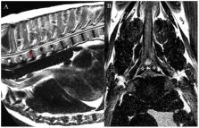
          The aim of the study was to describe the signalment, clinical presentation and presumptive or final diagnoses of dogs with cranial thoracic spinal cord lesions identified on advanced imaging. Retrospective evaluation of the databases of three veterinary specialty centres, between 2009 and 2021, was performed to identify dogs with a lesion affecting the cranial thoracic vertebral column (T1-T6 vertebrae) as the primary cause for presenting signs of myelopathy and/or spinal pain. Eighty-four dogs were included in the study, with the majority ( n = 76) presenting with a progressive history of over 4-weeks' duration. On neurologic examination, most dogs were ambulatory ( n = 64), and the most common neuroanatomic localisation was the T3-L3 spinal cord segments ( n = 63). Twelve dogs (14%) showed a short-strided thoracic limb gait on clinical examination. The most common diagnosis was neoplasia ( n = 33), followed by anomalies ( n = 22, including vertebral body malformations in 14 dogs) and degenerative disorders ( n = 16, with intervertebral disc protrusion diagnosed in 9 dogs). The most common vertebrae affected were T3 and T5. Most dogs with degenerative conditions showed asymmetric clinical signs, and the majority of dogs with neoplasia showed signs of spinal hyperaesthesia on examination. The findings of this study describe the clinical signs and presumptive or final diagnoses associated with lesions affecting the cranial thoracic spinal cord. When combined with the signalment and clinical history, this information can assist in both the recognition of and problem-based approach to these cases.

          Related collections

          Most cited references58

          • Record: found
          • Abstract: found
          • Article: not found

          Intervertebral disc disease in dogs.

          Intervertebral disc herniation is a common cause of neurologic dysfunction in dogs. This article reviews the anatomy, pathophysiology, diagnostic imaging, treatment options, and prognosis for canine cervical and thoracolumbar intervertebral disc disease. The extensive literature pertinent to intervertebral disc disease is reviewed with the goal of summarizing the information available to help clinicians in their decision making. Copyright 2010 Elsevier Inc. All rights reserved.
            Bookmark
            • Record: found
            • Abstract: found
            • Article: not found

            Congenital spinal malformations in small animals.

            Congenital anomalies of the spine are common in small animals. The type of deformity, location, severity, time of onset of associated clinical signs, and progression of neurologic dysfunction varies widely. To promote clearer understanding, the authors present the various spinal malformations using modified human classification schemes and use current widely accepted definitions and terminology. The diagnostic approach, including utilization of advanced imaging, and surgical management is emphasized. Copyright 2010. Published by Elsevier Inc.
              Bookmark
              • Record: found
              • Abstract: found
              • Article: not found

              Spinal neoplasms in small animals.

              Tumors arising from or involving the spinal cord are important considerations in animals presented for pain and limb dysfunction. Clinical signs of spinal cord dysfunction, however, are not pathognomic for neoplastic disease in most instances. Advanced magnetic resonance imaging (MRI) often accurately identifies the location and extent of abnormalities. Although some spinal neoplasms have a characteristic appearance with MRI, in other instances the abnormalities may not be readily discernable as neoplastic. Histologic diagnosis, therefore, is imperative to provide information regarding potential treatment modalities and prognosis. Histologic diagnosis is most commonly performed following surgical biopsy and is often performed in combination with surgical removal.
                Bookmark

                Author and article information

                Contributors
                Journal
                Front Vet Sci
                Front Vet Sci
                Front. Vet. Sci.
                Frontiers in Veterinary Science
                Frontiers Media S.A.
                2297-1769
                12 September 2022
                2022
                : 9
                : 960912
                Affiliations
                [1] 1Anderson Moores Veterinary Specialists, Part of Linnaeus Veterinary Limited , Hursley, United Kingdom
                [2] 2School of Veterinary Medicine, College of Medical, Veterinary and Life Sciences, University of Glasgow , Glasgow, United Kingdom
                [3] 3Department of Veterinary Medicine, University of Cambridge , Cambridge, United Kingdom
                [4] 4Departamento de Medicina y Cirugía Animal, Facultad de Veterinaria, Universidad Cardenal Herrera-CEU, CEU Universities , Valencia, Spain
                Author notes

                Edited by: Zoe Polizopoulou, Aristotle University of Thessaloniki, Greece

                Reviewed by: Rania Baka, Aristotle University of Thessaloniki, Greece; Christoforos Posporis, Pride Veterinary Centre, United Kingdom

                *Correspondence: Bruno A. Lopes bruno.lopes@ 123456linnaeusgroup.co.uk

                This article was submitted to Veterinary Neurology and Neurosurgery, a section of the journal Frontiers in Veterinary Science

                †Present address: Roberto José-López, Hamilton Specialist Referrals – IVC Evidensia, High Wycombe, United Kingdom

                Daniel Sánchez-Masián, Veterios Referral Hospital, Madrid, Spain

                Article
                10.3389/fvets.2022.960912
                9510707
                70459517-5ad7-4a7b-b520-7e581171a94b
                Copyright © 2022 Lopes, Ives, José-López, Gutierrez-Quintana, Abouzeid, Freeman, Redondo and Sánchez-Masián.

                This is an open-access article distributed under the terms of the Creative Commons Attribution License (CC BY). The use, distribution or reproduction in other forums is permitted, provided the original author(s) and the copyright owner(s) are credited and that the original publication in this journal is cited, in accordance with accepted academic practice. No use, distribution or reproduction is permitted which does not comply with these terms.

                History
                : 03 June 2022
                : 24 August 2022
                Page count
                Figures: 4, Tables: 3, Equations: 0, References: 58, Pages: 13, Words: 9718
                Categories
                Veterinary Science
                Original Research

                advanced imaging,ataxia,canine,neoplasia,neurological,spinal cord,two-engine gait

                Comments

                Comment on this article