7
views
0
recommends
+1 Recommend
0 collections
    0
    shares
      • Record: found
      • Abstract: found
      • Article: found
      Is Open Access

      Radiomics Analysis Based on Contrast-Enhanced MRI for Prediction of Therapeutic Response to Transarterial Chemoembolization in Hepatocellular Carcinoma

      research-article

      Read this article at

      Bookmark
          There is no author summary for this article yet. Authors can add summaries to their articles on ScienceOpen to make them more accessible to a non-specialist audience.

          Abstract

          Purpose

          To investigate the role of contrast-enhanced magnetic resonance imaging (CE-MRI) radiomics for pretherapeutic prediction of the response to transarterial chemoembolization (TACE) in patients with hepatocellular carcinoma (HCC).

          Methods

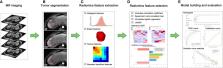
          One hundred and twenty-two HCC patients (objective response, n = 63; non-response, n = 59) who received CE-MRI examination before initial TACE were retrospectively recruited and randomly divided into a training cohort ( n = 85) and a validation cohort ( n = 37). All HCCs were manually segmented on arterial, venous and delayed phases of CE-MRI, and total 2367 radiomics features were extracted. Radiomics models were constructed based on each phase and their combination using logistic regression algorithm. A clinical-radiological model was built based on independent risk factors identified by univariate and multivariate logistic regression analyses. A combined model incorporating the radiomics score and selected clinical-radiological predictors was constructed, and the combined model was presented as a nomogram. Prediction models were evaluated by receiver operating characteristic curves, calibration curves, and decision curve analysis.

          Results

          Among all radiomics models, the three-phase radiomics model exhibited better performance in the training cohort with an area under the curve (AUC) of 0.838 (95% confidence interval (CI), 0.753 - 0.922), which was verified in the validation cohort (AUC, 0.833; 95% CI, 0.691 - 0.975). The combined model that integrated the three-phase radiomics score and clinical-radiological risk factors (total bilirubin, tumor shape, and tumor encapsulation) showed excellent calibration and predictive capability in the training and validation cohorts with AUCs of 0.878 (95% CI, 0.806 - 0.950) and 0.833 (95% CI, 0.687 - 0.979), respectively, and showed better predictive ability ( P = 0.003) compared with the clinical-radiological model (AUC, 0.744; 95% CI, 0.642 - 0.846) in the training cohort. A nomogram based on the combined model achieved good clinical utility in predicting the treatment efficacy of TACE.

          Conclusion

          CE-MRI radiomics analysis may serve as a promising and noninvasive tool to predict therapeutic response to TACE in HCC, which will facilitate the individualized follow-up and further therapeutic strategies guidance in HCC patients.

          Related collections

          Most cited references44

          • Record: found
          • Abstract: not found
          • Article: not found

          Hepatocellular Carcinoma

            Bookmark
            • Record: found
            • Abstract: not found
            • Article: not found

            Diagnosis, Staging, and Management of Hepatocellular Carcinoma: 2018 Practice Guidance by the American Association for the Study of Liver Diseases

              Bookmark
              • Record: found
              • Abstract: found
              • Article: not found

              Radiomics: extracting more information from medical images using advanced feature analysis.

              Solid cancers are spatially and temporally heterogeneous. This limits the use of invasive biopsy based molecular assays but gives huge potential for medical imaging, which has the ability to capture intra-tumoural heterogeneity in a non-invasive way. During the past decades, medical imaging innovations with new hardware, new imaging agents and standardised protocols, allows the field to move towards quantitative imaging. Therefore, also the development of automated and reproducible analysis methodologies to extract more information from image-based features is a requirement. Radiomics--the high-throughput extraction of large amounts of image features from radiographic images--addresses this problem and is one of the approaches that hold great promises but need further validation in multi-centric settings and in the laboratory. Copyright © 2011 Elsevier Ltd. All rights reserved.
                Bookmark

                Author and article information

                Contributors
                Journal
                Front Oncol
                Front Oncol
                Front. Oncol.
                Frontiers in Oncology
                Frontiers Media S.A.
                2234-943X
                31 March 2021
                2021
                : 11
                : 582788
                Affiliations
                [1] 1Department of Radiology, First Affiliated Hospital, Dalian Medical University , Dalian, China
                [2] 2Chengdu Institute of Computer Application, Chinese Academy of Sciences , Chengdu, China
                [3] 3University of Chinese Academy of Sciences , Beijing, China
                [4] 4Department of Interventional Radiology, First Affiliated Hospital, Dalian Medical University , Dalian, China
                [5] 5Life Sciences, GE Healthcare , Shanghai, China
                [6] 6Global Research, GE Healthcare , Shanghai, China
                [7] 7Clinical Education Team (CET), GE Healthcare , Shanghai, China
                Author notes

                Edited by: Wenli Cai, Massachusetts General Hospital and Harvard Medical School, United States

                Reviewed by: Haiyi Wang, Chinese People’s Liberation Army General Hospital, China; Feng Chen, Zhejiang Chinese Medical University, China

                *Correspondence: Ai Lian Liu, liuailian@ 123456dmu.edu.cn

                This article was submitted to Cancer Imaging and Image-directed Interventions, a section of the journal Frontiers in Oncology

                Article
                10.3389/fonc.2021.582788
                8045706
                33868988
                70053c87-2917-4fb9-af1e-8346306901e1
                Copyright © 2021 Zhao, Wang, Wu, Zhang, Lin, Yao, Chen, Wang, Sheng, Liu, Song, Wang, An, Guo, Li, Wu and Liu

                This is an open-access article distributed under the terms of the Creative Commons Attribution License (CC BY). The use, distribution or reproduction in other forums is permitted, provided the original author(s) and the copyright owner(s) are credited and that the original publication in this journal is cited, in accordance with accepted academic practice. No use, distribution or reproduction is permitted which does not comply with these terms.

                History
                : 13 July 2020
                : 09 March 2021
                Page count
                Figures: 6, Tables: 2, Equations: 0, References: 44, Pages: 12, Words: 6539
                Categories
                Oncology
                Original Research

                Oncology & Radiotherapy
                hepatocellular carcinoma,radiomics,magnetic resonance imaging,transarterial chemoembolization,therapeutic response

                Comments

                Comment on this article