13
views
0
recommends
+1 Recommend
0 collections
    0
    shares
      • Record: found
      • Abstract: found
      • Article: found
      Is Open Access

      Interplay between Müller cells and microglia aggravates retinal inflammatory response in experimental glaucoma

      research-article

      Read this article at

      Bookmark
          There is no author summary for this article yet. Authors can add summaries to their articles on ScienceOpen to make them more accessible to a non-specialist audience.

          Abstract

          Background

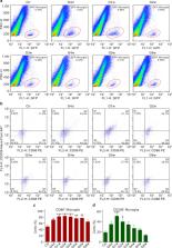
          Glaucoma, the leading cause of irreversible blindness, is a retinal neurodegenerative disease, which results from progressive apoptotic death of retinal ganglion cells (RGCs). Although the mechanisms underlying RGC apoptosis in glaucoma are extremely complicated, an abnormal cross-talk between retinal glial cells and RGCs is generally thought to be involved. However, how interaction of Müller cells and microglia, two types of glial cells, contributes to RGC injury is largely unknown.

          Methods

          A mouse chronic ocular hypertension (COH) experimental glaucoma model was produced. Western blotting, immunofluorescence, quantitative real-time polymerase chain reaction (q-PCR), transwell co-culture of glial cells, flow cytometry assay, ELISA, Ca 2+ image, and terminal deoxynucleotidyl transferase dUTP nick end labeling (TUNEL) techniques were employed to investigate the interaction of Müller cells and microglia, and its underlying mechanisms in COH retina.

          Results

          We first showed that Müller cell activation in mice with COH induced microglia activation through the ATP/P2X7 receptor pathway. The activation of microglia resulted in a significant increase in mRNA and protein levels of pro-inflammatory factors, such as tumor necrosis factor-α and interleukin-6. These inflammatory factors in turn caused the up-regulation of mRNA expression of pro-inflammatory factors in Müller cells through a positive feedback manner.

          Conclusions

          These findings provide robust evidence, for the first time, that retinal inflammatory response may be aggravated by an interplay between activated two types of glial cells. These results also suggest that to reduce the interplay between Müller cells and microglia could be a potential effective strategy for preventing the loss of RGCs in glaucoma.

          Supplementary Information

          The online version contains supplementary material available at 10.1186/s12974-021-02366-x.

          Related collections

          Most cited references51

          • Record: found
          • Abstract: found
          • Article: not found

          Differential Roles of M1 and M2 Microglia in Neurodegenerative Diseases.

          One of the most striking hallmarks shared by various neurodegenerative diseases, including Parkinson's disease, Alzheimer's disease (AD), and amyotrophic lateral sclerosis, is microglia-mediated neuroinflammation. Increasing evidence indicates that microglial activation in the central nervous system is heterogeneous, which can be categorized into two opposite types: M1 phenotype and M2 phenotype. Depending on the phenotypes activated, microglia can produce either cytotoxic or neuroprotective effects. In this review, we focus on the potential role of M1 and M2 microglia and the dynamic changes of M1/M2 phenotypes that are critically associated with the neurodegenerative diseases. Generally, M1 microglia predominate at the injury site at the end stage of disease, when the immunoresolution and repair process of M2 microglia are dampened. This phenotype transformation is very complicated in AD due to the phagocytosis of regionally distributed β-amyloid (Aβ) plaque and tangles that are released into the extracellular space. The endogenous stimuli including aggregated α-synuclein, mutated superoxide dismutase, Aβ, and tau oligomers exist in the milieu that may persistently activate M1 pro-inflammatory responses and finally lead to irreversible neuron loss. The changes of microglial phenotypes depend on the disease stages and severity; mastering the stage-specific switching of M1/M2 phenotypes within appropriate time windows may provide better therapeutic benefit.
            Bookmark
            • Record: found
            • Abstract: found
            • Article: not found

            A polarizing question: do M1 and M2 microglia exist?

            Microglial research has entered a fertile, dynamic phase characterized by novel technologies including two-photon imaging, whole-genome transcriptomic and epigenomic analysis with complementary bioinformatics, unbiased proteomics, cytometry by time of flight (CyTOF; Fluidigm) cytometry, and complex high-content experimental models including slice culture and zebrafish. Against this vivid background of newly emerging data, investigators will encounter in the microglial research literature a body of published work using the terminology of macrophage polarization, most commonly into the M1 and M2 phenotypes. It is the assertion of this opinion piece that microglial polarization has not been established by research findings. Rather, the adoption of this schema was undertaken in an attempt to simplify data interpretation at a time when the ontogeny and functional significance of microglia had not yet been characterized. Now, terminology suggesting established meaningful pathways of microglial polarization hinders rather than aids research progress and should be discarded.
              Bookmark
              • Record: found
              • Abstract: found
              • Article: not found

              Müller cells in the healthy and diseased retina.

              Müller glial cells span the entire thickness of the tissue, and ensheath all retinal neurons, in vertebrate retinae of all species. This morphological relationship is reflected by a multitude of functional interactions between neurons and Müller cells, including a 'metabolic symbiosis' and the processing of visual information. Müller cells are also responsible for the maintenance of the homeostasis of the retinal extracellular milieu (ions, water, neurotransmitter molecules, and pH). In vascularized retinae, Müller cells may also be involved in the control of angiogenesis, and the regulation of retinal blood flow. Virtually every disease of the retina is associated with a reactive Müller cell gliosis which, on the one hand, supports the survival of retinal neurons but, on the other hand, may accelerate the progress of neuronal degeneration: Müller cells protect neurons via a release of neurotrophic factors, the uptake and degradation of the excitotoxin, glutamate, and the secretion of the antioxidant, glutathione. However, gliotic Müller cells display a dysregulation of various neuron-supportive functions. This contributes to a disturbance of retinal glutamate metabolism and ion homeostasis, and causes the development of retinal edema and neuronal cell death. Moreover, there are diseases evoking a primary Müller cell insufficiency, such as hepatic retinopathy and certain forms of glaucoma. Any impairment of supportive functions of Müller cells, primary or secondary, must cause and/or aggravate a dysfunction and loss of neurons, by increasing the susceptibility of neurons to stressful stimuli in the diseased retina. On the contrary, Müller cells may be used in the future for novel therapeutic strategies to protect neurons against apoptosis (somatic gene therapy), or to differentiate retinal neurons from Müller/stem cells. Meanwhile, a proper understanding of the gliotic responses of Müller cells in the diseased retina, and of their protective vs. detrimental effects, is essential for the development of efficient therapeutic strategies that use and stimulate the neuron-supportive/protective-and prevent the destructive-mechanisms of gliosis.
                Bookmark

                Author and article information

                Contributors
                17111520019@fudan.edu.cn
                19111520035@fudan.edu.cn
                18111520030@fudan.edu.cn
                19111520010@fudan.edu.cn
                fanglee@fudan.edu.cn
                yymiao@fudan.edu.cn
                bolei99@126.com
                xlyang@fudan.edu.cn
                zfwang@fudan.edu.cn
                Journal
                J Neuroinflammation
                J Neuroinflammation
                Journal of Neuroinflammation
                BioMed Central (London )
                1742-2094
                24 December 2021
                24 December 2021
                2021
                : 18
                : 303
                Affiliations
                [1 ]GRID grid.8547.e, ISNI 0000 0001 0125 2443, State Key Laboratory of Medical Neurobiology and MOE Frontiers Center for Brain Science, Institutes of Brain Science, , Fudan University, ; Shanghai, 200032 China
                [2 ]GRID grid.207374.5, ISNI 0000 0001 2189 3846, Institute of Neuroscience and Third Affiliated Hospital, Henan Provincial People’s Hospital, Henan Eye Institute, Henan Eye Hospital, People’s Hospital of Zhengzhou University, , Zhengzhou University, ; Zhengzhou, 450003 China
                Author information
                http://orcid.org/0000-0002-2279-6885
                Article
                2366
                10.1186/s12974-021-02366-x
                8705189
                34952606
                6ff75af2-f0ef-4a73-b468-b715e981a52a
                © The Author(s) 2021

                Open AccessThis article is licensed under a Creative Commons Attribution 4.0 International License, which permits use, sharing, adaptation, distribution and reproduction in any medium or format, as long as you give appropriate credit to the original author(s) and the source, provide a link to the Creative Commons licence, and indicate if changes were made. The images or other third party material in this article are included in the article's Creative Commons licence, unless indicated otherwise in a credit line to the material. If material is not included in the article's Creative Commons licence and your intended use is not permitted by statutory regulation or exceeds the permitted use, you will need to obtain permission directly from the copyright holder. To view a copy of this licence, visit http://creativecommons.org/licenses/by/4.0/. The Creative Commons Public Domain Dedication waiver ( http://creativecommons.org/publicdomain/zero/1.0/) applies to the data made available in this article, unless otherwise stated in a credit line to the data.

                History
                : 14 September 2021
                : 18 December 2021
                Funding
                Funded by: FundRef http://dx.doi.org/10.13039/501100001809, National Natural Science Foundation of China;
                Award ID: 81790642
                Award Recipient :
                Funded by: FundRef http://dx.doi.org/10.13039/501100010229, Natural Science Foundation of Tianjin Municipal Science and Technology Commission;
                Award ID: 31872765
                Award Recipient :
                Categories
                Research
                Custom metadata
                © The Author(s) 2021

                Neurosciences
                glaucoma,müller cells,microglia,activation,inflammatory response,atp/p2x7 receptor
                Neurosciences
                glaucoma, müller cells, microglia, activation, inflammatory response, atp/p2x7 receptor

                Comments

                Comment on this article