0
views
0
recommends
+1 Recommend
0 collections
    0
    shares
      • Record: found
      • Abstract: found
      • Article: found
      Is Open Access

      Agreement, repeatability, and reproducibility of quantitative retinal layer assessment using swept-source and spectral-domain optical coherence tomography in eyes with retinal diseases

      research-article

      Read this article at

      Bookmark
          There is no author summary for this article yet. Authors can add summaries to their articles on ScienceOpen to make them more accessible to a non-specialist audience.

          Abstract

          Purpose

          To evaluate the agreement and precision of retinal thickness measurements obtained using swept-source optical coherence tomography (SS-OCT) and spectral-domain OCT (SD-OCT) in healthy eyes and eyes with retinopathy.

          Methods

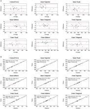
          This cross-sectional prospective study involved three DRI-OCT Triton (SS-OCT) and three 3D-OCT-1 Maestro (SD-OCT) devices. One of each device (Maestro and Triton) was paired with a single operator. Healthy subjects and patients with retinal diseases were recruited, with study eye and testing order randomized. At least 3 scans per eye were captured for wide scan (12 mm × 9 mm-Triton and Maestro) and macular cube scan (7 mm × 7 mm-Triton, 6 mm × 6 mm-Maestro). Thickness of the full retina, ganglion cell layer + inner plexiform layer (GCL+), and ganglion cell complex (GCL++) were obtained from wide scan and cube scans. Agreement of the measurements between the Triton and Maestro was evaluated by Bland–Altman analysis and Deming regression for each group. Repeatability and reproducibility were assessed using a two-way random effect analysis of variance (ANOVA) model for each parameter by group.

          Results

          Twenty-five healthy subjects (25 eyes) and 26 patients with retinal diseases (26 eyes), including, but not limited to, age-related macular degeneration, macular hole, and diabetic retinopathy were recruited. Overall, the measurement differences between Triton and Maestro were <6 μm (mean differences of full retina, GCL++, and GCL+ thickness were ≤5.5 μm, 1.3 μm, and 2.8 μm, respectively) and not statistically significant across the parameters. The repeatability and reproducibility estimates indicate high precision in both devices and groups. Across all the parameters, the repeatability limit was ≤7.6 μm for Triton and ≤12.7 μm for Maestro; reproducibility limit was ≤9.2 μm for Triton and ≤14.4 μm for Maestro. In eyes with retinal pathology, the repeatability coefficient of variation (CV)% was ≤2.6% for Triton and ≤3.4% for Maestro; reproducibility CV% was ≤3.3% for Triton and ≤3.5% for Maestro.

          Conclusion

          Both Triton SS-OCT and Maestro SD-OCT provide reliable measurements of retinal thickness in healthy eyes and eyes with retinal diseases. Excellent agreement between the two devices indicates interoperability when testing healthy eyes or eyes with retinal pathology. These findings support the use of thickness measurements from Triton SS-OCT and Maestro SD-OCT in clinical practice.

          Related collections

          Most cited references29

          • Record: found
          • Abstract: found
          • Article: not found

          Optical coherence tomography--current and future applications.

          Optical coherence tomography (OCT) has revolutionized the clinical practice of ophthalmology. It is a noninvasive imaging technique that provides high-resolution, cross-sectional images of the retina, retinal nerve fiber layer and the optic nerve head. This review discusses the present applications of the commercially available spectral-domain OCT (SD-OCT) systems in the diagnosis and management of retinal diseases, with particular emphasis on choroidal imaging. Future directions of OCT technology and their potential clinical uses are discussed. Analysis of the choroidal thickness in healthy eyes and disease states such as age-related macular degeneration, central serous chorioretinopathy, diabetic retinopathy and inherited retinal dystrophies has been successfully achieved using SD-OCT devices with software improvements. Future OCT innovations such as longer-wavelength OCT systems including the swept-source technology, along with Doppler OCT and en-face imaging, may improve the detection of subtle microstructural changes in chorioretinal diseases by improving imaging of the choroid. Advances in OCT technology provide for better understanding of pathogenesis, improved monitoring of progression and assistance in quantifying response to treatment modalities in diseases of the posterior segment of the eye. Further improvements in both hardware and software technologies should further advance the clinician's ability to assess and manage chorioretinal diseases.
            Bookmark
            • Record: found
            • Abstract: found
            • Article: not found

            Optical coherence tomography: imaging of the choroid and beyond.

            Seventy percent of the blood flow to the eye goes to the choroid, a structure that is vitally important to the function of the retina. The in vivo structure of the choroid in health and disease is incompletely visualized with traditional imaging modalities, including indocyanine green angiography, ultrasonography, and spectral domain optical coherence tomography (OCT). Use of new OCT modalities, including enhanced depth imaging OCT, image averaging, and swept-source OCT, have led to increased visualization of the choroidal anatomy. The correlation of these new anatomical findings with other imaging modalities results increases understanding of many eye diseases and recognises of new ones. The status of the choroid appears to be a crucial determinant in the pathogenesis of diseases such as age-related choroidal atrophy, myopic chorioretinal atrophy, central serous chorioretinopathy, chorioretinal inflammatory diseases, and tumors. Extension of these imaging techniques has provided insights into abnormalities of the sclera and optic nerve. Future developments will include blood flow information, 3D rendering of various ocular structures, and the ability to evaluate changes in 3D structural information over time (4D imaging). Copyright © 2013 Elsevier Inc. All rights reserved.
              Bookmark
              • Record: found
              • Abstract: found
              • Article: not found

              Retinal thickness decreases with age: an OCT study.

              In three dimensional optic disc tomography a reference plane is required to calculate optic disc rim or cup values. The position of the reference plane often depends on the retinal thickness at the temporal disc margin. Originally it was assumed that the retinal thickness at the temporal disc margin is independent of age. The aim of the study was to check this hypothesis using optical coherence tomography, and additionally to determine the reproducibility of OCT measurements in this area. 100 eyes of 100 healthy volunteers were included in this study. Three OCT scans were performed on each eye. The scans were aligned vertically and placed at the temporal edge of the optic disc. For each eye, the thickness of the whole retina as well as the thickness of the retinal nerve fibre layer were calculated together with their coefficients of variation. Thereafter retinal thickness and nerve fibre layer thickness were correlated with age. The mean retinal thickness was 249 (SD 22) micro m. The mean nerve fibre layer thickness was 109 (22) micro m. The mean coefficients of variation were 3.5% (total retinal thickness) and 8.0% (nerve fibre layer thickness). Both the total retinal thickness and the nerve fibre layer thickness were significantly correlated with age (total retina: y = 269.5 - 0.53 x x; R(2) = 0.133; p = 0.0002, nerve fibre layer: y = 126.8 - 0.44 x x; R(2) = 0.094; p<0.0019. Using OCT scans the total retinal thickness can be calculated with high reproducibility (coefficient of variation = 3.5%). The reproducibility of nerve fibre layer thickness measurements is clearly lower (coefficient of variation = 8.0%). Both the total retinal thickness and the nerve fibre layer thickness significantly decrease with age. This influence of the age related decrease in RNFL/retinal thickness on the reference plane, however, is negligible.
                Bookmark

                Author and article information

                Contributors
                URI : https://loop.frontiersin.org/people/1163811/overviewRole: Role: Role: Role: Role: Role: Role:
                URI : https://loop.frontiersin.org/people/2537685/overviewRole: Role: Role: Role: Role: Role: Role: Role: Role:
                URI : https://loop.frontiersin.org/people/2579623/overviewRole: Role: Role:
                Role: Role: Role: Role:
                Role: Role: Role:
                Journal
                Front Med (Lausanne)
                Front Med (Lausanne)
                Front. Med.
                Frontiers in Medicine
                Frontiers Media S.A.
                2296-858X
                18 December 2023
                2023
                : 10
                : 1281751
                Affiliations
                [1] 1Topcon Healthcare , Oakland, NJ, United States
                [2] 2Fischer Laser Eye Center , Willmar, MN, United States
                [3] 3Doheny Eye Institute , Pasadena, CA, United States
                [4] 4Department of Ophthalmology, David Geffen School of Medicine, University of California, Los Angeles , Los Angeles, CA, United States
                Author notes

                Edited by: Solmaz Abdolrahimzadeh, Sapienza University of Rome, Italy

                Reviewed by: Mengxi Shen, University of Miami Health System, United States; Ana Rita Santos, Association for Innovation and Biomedical Research on Light and Image (AIBILI), Portugal

                *Correspondence: Huiyuan Hou, hhou@ 123456topcon.com
                Article
                10.3389/fmed.2023.1281751
                10757970
                38164223
                6fb77d4b-4c51-44d1-9636-d9c46de74616
                Copyright © 2023 Hou, Durbin, El-Nimri, Fischer and Sadda.

                This is an open-access article distributed under the terms of the Creative Commons Attribution License (CC BY). The use, distribution or reproduction in other forums is permitted, provided the original author(s) and the copyright owner(s) are credited and that the original publication in this journal is cited, in accordance with accepted academic practice. No use, distribution or reproduction is permitted which does not comply with these terms.

                History
                : 23 August 2023
                : 04 December 2023
                Page count
                Figures: 2, Tables: 5, Equations: 0, References: 29, Pages: 13, Words: 7616
                Funding
                The author(s) declare that no financial support was received for the research, authorship, and/or publication of this article.
                Categories
                Medicine
                Original Research
                Custom metadata
                Ophthalmology

                optical coherence tomography,repeatability,reproducibility,agreement,retinopathy

                Comments

                Comment on this article