6
views
0
recommends
+1 Recommend
0 collections
    0
    shares
      • Record: found
      • Abstract: found
      • Article: found
      Is Open Access

      Chronic exposure to tramadol induces cardiac inflammation and endothelial dysfunction in mice

      research-article

      Read this article at

      Bookmark
          There is no author summary for this article yet. Authors can add summaries to their articles on ScienceOpen to make them more accessible to a non-specialist audience.

          Abstract

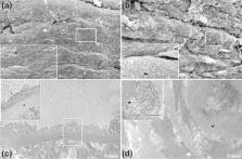
          Tramadol is an opioid extensively used to treat moderate to severe pain; however, prolonged therapy is associated with several tissues damage. Chronic use of tramadol was linked to increased hospitalizations due to cardiovascular complications. Limited literature has described the effects of tramadol on the cardiovascular system, so we sought to investigate these actions and elucidate the underlying mechanisms. Mice received tramadol hydrochloride (40 mg/kg body weight) orally for 4 successive weeks. Oxidative stress, inflammation, and cardiac toxicity were assessed. In addition, eNOS expression was evaluated. Our results demonstrated marked histopathological alteration in heart and aortic tissues after exposure to tramadol. Tramadol upregulated the expression of oxidative stress and inflammatory markers in mice heart and aorta, whereas downregulated eNOS expression. Tramadol caused cardiac damage shown by the increase in LDH, Troponin I, and CK-MB activities in serum samples. Overall, these results highlight the risks of tramadol on the cardiovascular system.

          Related collections

          Most cited references74

          • Record: found
          • Abstract: found
          • Article: not found

          G*Power 3: A flexible statistical power analysis program for the social, behavioral, and biomedical sciences

          G*Power (Erdfelder, Faul, & Buchner, 1996) was designed as a general stand-alone power analysis program for statistical tests commonly used in social and behavioral research. G*Power 3 is a major extension of, and improvement over, the previous versions. It runs on widely used computer platforms (i.e., Windows XP, Windows Vista, and Mac OS X 10.4) and covers many different statistical tests of the t, F, and chi2 test families. In addition, it includes power analyses for z tests and some exact tests. G*Power 3 provides improved effect size calculators and graphic options, supports both distribution-based and design-based input modes, and offers all types of power analyses in which users might be interested. Like its predecessors, G*Power 3 is free.
            Bookmark
            • Record: found
            • Abstract: found
            • Article: found
            Is Open Access

            Lipid Peroxidation: Production, Metabolism, and Signaling Mechanisms of Malondialdehyde and 4-Hydroxy-2-Nonenal

            Lipid peroxidation can be described generally as a process under which oxidants such as free radicals attack lipids containing carbon-carbon double bond(s), especially polyunsaturated fatty acids (PUFAs). Over the last four decades, an extensive body of literature regarding lipid peroxidation has shown its important role in cell biology and human health. Since the early 1970s, the total published research articles on the topic of lipid peroxidation was 98 (1970–1974) and has been increasing at almost 135-fold, by up to 13165 in last 4 years (2010–2013). New discoveries about the involvement in cellular physiology and pathology, as well as the control of lipid peroxidation, continue to emerge every day. Given the enormity of this field, this review focuses on biochemical concepts of lipid peroxidation, production, metabolism, and signaling mechanisms of two main omega-6 fatty acids lipid peroxidation products: malondialdehyde (MDA) and, in particular, 4-hydroxy-2-nonenal (4-HNE), summarizing not only its physiological and protective function as signaling molecule stimulating gene expression and cell survival, but also its cytotoxic role inhibiting gene expression and promoting cell death. Finally, overviews of in vivo mammalian model systems used to study the lipid peroxidation process, and common pathological processes linked to MDA and 4-HNE are shown.
              Bookmark
              • Record: found
              • Abstract: found
              • Article: not found

              Endothelial nitric oxide synthase in vascular disease: from marvel to menace.

              Nitric oxide (NO*) is an important protective molecule in the vasculature, and endothelial NO* synthase (eNOS) is responsible for most of the vascular NO* produced. A functional eNOS oxidizes its substrate L-arginine to L-citrulline and NO*. This normal function of eNOS requires dimerization of the enzyme, the presence of the substrate L-arginine, and the essential cofactor (6R)-5,6,7,8-tetrahydro-L-biopterin (BH4), one of the most potent naturally occurring reducing agents. Cardiovascular risk factors such as hypertension, hypercholesterolemia, diabetes mellitus, or chronic smoking stimulate the production of reactive oxygen species in the vascular wall. Nicotinamide adenine dinucleotide phosphate (NADPH) oxidases represent major sources of this reactive oxygen species and have been found upregulated and activated in animal models of hypertension, diabetes, and sedentary lifestyle and in patients with cardiovascular risk factors. Superoxide (O2*-) reacts avidly with vascular NO* to form peroxynitrite (ONOO-). The cofactor BH4 is highly sensitive to oxidation by ONOO-. Diminished levels of BH4 promote O2*- production by eNOS (referred to as eNOS uncoupling). This transformation of eNOS from a protective enzyme to a contributor to oxidative stress has been observed in several in vitro models, in animal models of cardiovascular diseases, and in patients with cardiovascular risk factors. In many cases, supplementation with BH4 has been shown to correct eNOS dysfunction in animal models and patients. In addition, folic acid and infusions of vitamin C are able to restore eNOS functionality, most probably by enhancing BH4 levels as well.
                Bookmark

                Author and article information

                Contributors
                m.elbadawy@aun.edu.eg
                Journal
                Sci Rep
                Sci Rep
                Scientific Reports
                Nature Publishing Group UK (London )
                2045-2322
                21 September 2021
                21 September 2021
                2021
                : 11
                : 18772
                Affiliations
                [1 ]GRID grid.252487.e, ISNI 0000 0000 8632 679X, Department of Histology and Cell Biology, Faculty of Medicine, , Assiut University, ; Assiut, 71515 Egypt
                [2 ]GRID grid.252487.e, ISNI 0000 0000 8632 679X, Department of Medical Biochemistry, Faculty of Medicine, , Assiut University, ; Assiut, Egypt
                [3 ]GRID grid.252487.e, ISNI 0000 0000 8632 679X, Department of Microbiology and Immunology, Faculty of Medicine, , Assiut University, ; Assiut, Egypt
                [4 ]GRID grid.252487.e, ISNI 0000 0000 8632 679X, Department of Human Anatomy and Embryology, Faculty of Medicine, , Assiut University, ; Assiut, Egypt
                [5 ]GRID grid.412602.3, ISNI 0000 0000 9421 8094, Department of Basic Medical Sciences, Unaizah College of Medicine and Medical Sciences, , Qassim University, ; Unaizah, Kingdom of Saudi Arabia
                [6 ]GRID grid.494608.7, ISNI 0000 0004 6027 4126, Department of Anatomy, College of Medicine, , Bisha University, ; Bisha, Kingdom of Saudi Arabia
                [7 ]Department of Biochemistry, Sphinx University, Assiut, Egypt
                Author information
                http://orcid.org/0000-0001-8833-7916
                Article
                98206
                10.1038/s41598-021-98206-2
                8455605
                34548593
                6f63c03e-c8e8-474b-bc7d-e070532ca7f5
                © The Author(s) 2021

                Open Access This article is licensed under a Creative Commons Attribution 4.0 International License, which permits use, sharing, adaptation, distribution and reproduction in any medium or format, as long as you give appropriate credit to the original author(s) and the source, provide a link to the Creative Commons licence, and indicate if changes were made. The images or other third party material in this article are included in the article's Creative Commons licence, unless indicated otherwise in a credit line to the material. If material is not included in the article's Creative Commons licence and your intended use is not permitted by statutory regulation or exceeds the permitted use, you will need to obtain permission directly from the copyright holder. To view a copy of this licence, visit http://creativecommons.org/licenses/by/4.0/.

                History
                : 22 March 2021
                : 6 September 2021
                Categories
                Article
                Custom metadata
                © The Author(s) 2021

                Uncategorized
                biochemistry,cell biology,electron microscopy
                Uncategorized
                biochemistry, cell biology, electron microscopy

                Comments

                Comment on this article