23
views
0
recommends
+1 Recommend
0 collections
    0
    shares
      • Record: found
      • Abstract: found
      • Article: found
      Is Open Access

      TFE3-associated perivascular epithelioid cell tumor with complete response to mTOR inhibitor therapy: report of first case and literature review

      case-report

      Read this article at

      Bookmark
          There is no author summary for this article yet. Authors can add summaries to their articles on ScienceOpen to make them more accessible to a non-specialist audience.

          Abstract

          Background

          Perivascular epitheloid cell tumor (PEComas) are characterized by expression of both muscles, most often smooth muscle actin (in ~80% of cases) and melanocytic markers (mainly HMB-45 and Melan A). TFE 3-associated PEComas are new variant which are poorly defined due to their limited reports in literature. These tumors lack response to targeted mTOR inhibitor therapy due to lack of mutation in TSC gene. Hereby, we are reporting a case of TFE3 associated pelvic PEComa showing excellent response to Everolimus.

          Case presentation

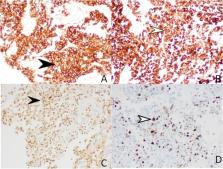
          A 45-year-old female presented with complaint of abdominal mass and bleeding per vaginum for 4 months. She had a history of total abdominal hysterectomy 3 years back in view of abnormal uterine bleeding and exploratory laprotomy 7 months back to remove some pelvic mass. Imaging suggested of ill-defined heterogenous mass of 9.3 x 9.2 x 16 cm involving the uterus, cervix, and upper 1/3 vagina. Multiple omental and peritoneal deposits were also seen, making probable diagnosis of carcinoma endometrium. USG guided biopsy showed cores of fibrous tissue with the presence of cells in sheets with granular eosinophillic cytoplasm; IHC showed positivity for TFE-3, H Caldesmon, GATA-3, and Melan A- and HMB-45; and Ki 67 index was 35%. The basis of above diagnosis of PEComa was made and she was started on Everolimus; repeat imaging after 3 months of therapy showed complete response.

          Conclusion

          We are reporting first case of malignant pelvic TFE 3 PEComa showing response to mTOR therapy. Identification of TFE 3 PEComa is important because they showed different biologic behavior then their conventional PEComa.

          Related collections

          Most cited references19

          • Record: found
          • Abstract: found
          • Article: not found

          Integrative genomic analyses identify MITF as a lineage survival oncogene amplified in malignant melanoma.

          Systematic analyses of cancer genomes promise to unveil patterns of genetic alterations linked to the genesis and spread of human cancers. High-density single-nucleotide polymorphism (SNP) arrays enable detailed and genome-wide identification of both loss-of-heterozygosity events and copy-number alterations in cancer. Here, by integrating SNP array-based genetic maps with gene expression signatures derived from NCI60 cell lines, we identified the melanocyte master regulator MITF (microphthalmia-associated transcription factor) as the target of a novel melanoma amplification. We found that MITF amplification was more prevalent in metastatic disease and correlated with decreased overall patient survival. BRAF mutation and p16 inactivation accompanied MITF amplification in melanoma cell lines. Ectopic MITF expression in conjunction with the BRAF(V600E) mutant transformed primary human melanocytes, and thus MITF can function as a melanoma oncogene. Reduction of MITF activity sensitizes melanoma cells to chemotherapeutic agents. Targeting MITF in combination with BRAF or cyclin-dependent kinase inhibitors may offer a rational therapeutic avenue into melanoma, a highly chemotherapy-resistant neoplasm. Together, these data suggest that MITF represents a distinct class of 'lineage survival' or 'lineage addiction' oncogenes required for both tissue-specific cancer development and tumour progression.
            Bookmark
            • Record: found
            • Abstract: found
            • Article: not found

            Perivascular epithelioid cell neoplasms of soft tissue and gynecologic origin: a clinicopathologic study of 26 cases and review of the literature.

            PEComas, occasionally associated with the tuberous sclerosis complex, are defined by the presence of perivascular epithelioid cells that coexpress muscle and melanocytic markers. This family of tumors includes angiomyolipoma (AML), clear cell sugar tumor of the lung (CCST), lymphangioleiomyomatosis (LAM), and very rare tumors in other locations. Because non-AML/non-LAM PEComas are extremely rare and their natural history and prognostic features undefined, we present our experience with 26 PEComas of soft tissue and the gynecologic tract, the largest series to date. We also performed a detailed review of the literature, with special attention to features predictive of clinical behavior. All PEComas exclusive of AML and LAM were retrieved from our consultation files. Immunohistochemistry for pan-cytokeratin (CK), S-100 protein, smooth muscle actins (SMA), desmin, vimentin, HMB45, Melan-A, microphthalmia transcription factor (MiTF), TFE3, CD117, and CD34 was performed. Clinical follow-up information was obtained. Fisher's exact test was performed. The median patient age was 46 years (range, 15-97 years); there was a marked female predominance (22 females, 4 males). Sites of involvement included the omentum or mesentery (6 cases), uterus (4 cases), pelvic soft tissues (3 cases), abdominal wall (2 cases), uterine cervix (2 cases), and vagina, retroperitoneum, thigh, falciform ligament, scalp, broad ligament, forearm, shoulder, and neck (1 case each). The tumors ranged from 1.6 to 29 cm in size (median, 7.8 cm). Tumors were epithelioid (N = 9), spindled (N = 7), or mixed (N = 10). Multinucleated giant cells were present in 18 cases. High nuclear grade was noted in 10 cases, high cellularity in 7 cases, necrosis in 8 cases, and vascular invasion in 3 cases. Mitotic activity was 0 to 50 mitotic figures (MF)/50 high power fields (HPF) (median, 0 MF/50 HPF) with atypical MF in 6 cases. IHC results were: SMA (20/25), desmin (8/22), HMB45 (22/24), Melan-A (13/18), MITF (9/18), S-100 protein (8/24), CK (3/23), vimentin (12/14), TFE3 (5/17), c-kit (1/20), and CD34 (0/7). Clinical follow-up (24 of 26 patients, 92%; median, 30 months; range, 10-84 months) showed 3 local recurrences and 5 distant metastases. At last available clinical follow-up, 2 patients (8%) were dead of disease, 4 patients (17%) were alive with metastatic or unresectable local disease, and 18 patients (75%) were alive with no evidence of disease. No patient in our series had a history of tuberous sclerosis complex. Recurrence and/or metastasis was strongly associated tumor size > median size (8 cm), mitotic activity greater than 1/50 HPF, and necrosis. We conclude that PEComas of soft tissue and gynecologic origin may be classified as "benign," "of uncertain malignant potential," or "malignant." Small PEComas without any worrisome histologic features are most likely benign. PEComas with nuclear pleomorphism alone ("symplastic") and large PEComas without other worrisome features have uncertain malignant potential. PEComas with two or more worrisome histologic features should be considered malignant. Occasional PEComas express unusual markers, such as S-100 protein, desmin, and rarely CK. The role of TFE3 in PEComas should be further studied.
              Bookmark
              • Record: found
              • Abstract: found
              • Article: not found

              Uterine PEComas: A Morphologic, Immunohistochemical, and Molecular Analysis of 32 Tumors

              Uterine perivascular epithelioid cell tumors (PEComas) are rare neoplasms that may show overlapping morphology and immunohistochemistry with uterine smooth muscle tumors. In this study, we evaluated the morphological, immunohistochemical, and molecular features of 32 PEComas, including 11 with aggressive behavior. Two distinct morphologies were observed: classic (n=30) and those with a lymphangioleiomyomatosis appearance (n=2). In the former, patients ranged from 32 to 77 (mean 51) years and 13% had tuberous sclerosis. Tumors ranged from 0.2 to 17 (mean 5.5) cm with 77% arising in the corpus. Epithelioid cells were present in 100% and a spindled component was seen in 37%. Nuclear atypia was low (53%), intermediate (17%), or high (30%). Mitoses ranged from 0–36 (mean 6) and 0–133 (mean 19) per 10 and 50 high-power fields, with atypical mitoses present in 30%. Thin and delicate vessels were noted in 100%, clear/eosinophilic and granular cytoplasm in 93%, stromal hyalinization in 73%, necrosis in 30%, and lymphovascular invasion in 10%. All tumors were positive for HMB-45, cathepsin K, and at least one muscle marker, with most expressing melan-A (77%) and/or MiTF (79%). A PSF-TFE3 fusion was identified in one while another showed a RAD51B-OPHN1 fusion. Follow-up ranged from two to 175 (mean 41) months, with 63% of patients alive and well, 20% dead of disease, 13% alive with disease, and 3% dead from other causes. In the latter, patients were 39 and 49 years old, one had tuberous sclerosis, while the other had pulmonary lymphangioleiomyomatosis. Both tumors expressed HMB-45, cathepsin K, and muscle markers, but lacked TFE3 and RAD51B rearrangements. The two patients are currently alive and well. Application of gynecologic-specific criteria (≥ 4 features required for malignancy: size ≥ 5 cm, high-grade atypia, mitoses > 1/50 high-power fields, necrosis, and lymphovascular invasion) for predicting outcome misclassified 36% (4/11) of aggressive tumors; thus, a modified algorithm with a threshold of three of these features is recommended to classify a PEComa as malignant.
                Bookmark

                Author and article information

                Contributors
                purwarroli@gmail.com
                Journal
                World J Surg Oncol
                World J Surg Oncol
                World Journal of Surgical Oncology
                BioMed Central (London )
                1477-7819
                1 March 2022
                1 March 2022
                2022
                : 20
                : 62
                Affiliations
                [1 ]GRID grid.411507.6, ISNI 0000 0001 2287 8816, Department of Surgical Oncology, Institute of Medical Sciences, , Banaras Hindu University, ; Varanasi, India
                [2 ]Department of Histopathology, Lal path labs, Varanasi, India
                [3 ]GRID grid.411507.6, ISNI 0000 0001 2287 8816, Department of Radiology and Imaging, Institute of Medical Sciences, , Banaras Hindu University, ; Varanasi, India
                Author information
                http://orcid.org/0000-0003-1245-239X
                Article
                2462
                10.1186/s12957-021-02462-5
                8886828
                35232443
                6ed7c66c-c1a7-4167-9878-e67849445d5a
                © The Author(s) 2022

                Open AccessThis article is licensed under a Creative Commons Attribution 4.0 International License, which permits use, sharing, adaptation, distribution and reproduction in any medium or format, as long as you give appropriate credit to the original author(s) and the source, provide a link to the Creative Commons licence, and indicate if changes were made. The images or other third party material in this article are included in the article's Creative Commons licence, unless indicated otherwise in a credit line to the material. If material is not included in the article's Creative Commons licence and your intended use is not permitted by statutory regulation or exceeds the permitted use, you will need to obtain permission directly from the copyright holder. To view a copy of this licence, visit http://creativecommons.org/licenses/by/4.0/. The Creative Commons Public Domain Dedication waiver ( http://creativecommons.org/publicdomain/zero/1.0/) applies to the data made available in this article, unless otherwise stated in a credit line to the data.

                History
                : 17 September 2021
                : 23 November 2021
                Categories
                Case Report
                Custom metadata
                © The Author(s) 2022

                Surgery
                perivascular epitheliod cell tumor,mtor therapy,tfe3,pecoma,uterus,gynecological
                Surgery
                perivascular epitheliod cell tumor, mtor therapy, tfe3, pecoma, uterus, gynecological

                Comments

                Comment on this article

                scite_
                0
                0
                0
                0
                Smart Citations
                0
                0
                0
                0
                Citing PublicationsSupportingMentioningContrasting
                View Citations

                See how this article has been cited at scite.ai

                scite shows how a scientific paper has been cited by providing the context of the citation, a classification describing whether it supports, mentions, or contrasts the cited claim, and a label indicating in which section the citation was made.

                Similar content499

                Cited by7

                Most referenced authors263