0
views
0
recommends
+1 Recommend
0 collections
    0
    shares
      • Record: found
      • Abstract: found
      • Article: found
      Is Open Access

      Deep Learning for Fully Automated Radiographic Measurements of the Pelvis and Hip

      Read this article at

      Bookmark
          There is no author summary for this article yet. Authors can add summaries to their articles on ScienceOpen to make them more accessible to a non-specialist audience.

          Abstract

          The morphometry of the hip and pelvis can be evaluated in native radiographs. Artificial-intelligence-assisted analyses provide objective, accurate, and reproducible results. This study investigates the performance of an artificial intelligence (AI)-based software using deep learning algorithms to measure radiological parameters that identify femoroacetabular impingement and hip dysplasia. Sixty-two radiographs (124 hips) were manually evaluated by three observers and fully automated analyses were performed by an AI-driven software (HIPPO™, ImageBiopsy Lab, Vienna, Austria). We compared the performance of the three human readers with the HIPPO™ using a Bayesian mixed model. For this purpose, we used the absolute deviation from the median ratings of all readers and HIPPO™. Our results indicate a high probability that the AI-driven software ranks better than at least one manual reader for the majority of outcome measures. Hence, fully automated analyses could provide reproducible results and facilitate identifying radiographic signs of hip disorders.

          Related collections

          Most cited references26

          • Record: found
          • Abstract: found
          • Article: not found

          Femoroacetabular impingement: a cause for osteoarthritis of the hip.

          A multitude of factors including biochemical, genetic, and acquired abnormalities may contribute to osteoarthritis of the hip. Although the pathomechanism of degenerative process affecting the dysplastic hip is well understood, the exact pathogenesis for idiopathic osteoarthritis has not been established. Based on clinical experience, with more than 600 surgical dislocations of the hip, allowing in situ inspection of the damage pattern and the dynamic proof of its origin, we propose femoroacetabular impingement as a mechanism for the development of early osteoarthritis for most nondysplastic hips. The concept focuses more on motion than on axial loading of the hip. Distinct clinical, radiographic, and intraoperative parameters can be used to confirm the diagnosis of this entity with timely delivery of treatment. Surgical treatment of femoroacetabular impingement focuses on improving the clearance for hip motion and alleviation of femoral abutment against the acetabular rim. It is proposed that early surgical intervention for treatment of femoroacetabular impingement, besides providing relief of symptoms, may decelerate the progression of the degenerative process for this group of young patients.
            Bookmark
            • Record: found
            • Abstract: found
            • Article: found
            Is Open Access

            Artificial intelligence versus clinicians: systematic review of design, reporting standards, and claims of deep learning studies

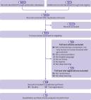
            Abstract Objective To systematically examine the design, reporting standards, risk of bias, and claims of studies comparing the performance of diagnostic deep learning algorithms for medical imaging with that of expert clinicians. Design Systematic review. Data sources Medline, Embase, Cochrane Central Register of Controlled Trials, and the World Health Organization trial registry from 2010 to June 2019. Eligibility criteria for selecting studies Randomised trial registrations and non-randomised studies comparing the performance of a deep learning algorithm in medical imaging with a contemporary group of one or more expert clinicians. Medical imaging has seen a growing interest in deep learning research. The main distinguishing feature of convolutional neural networks (CNNs) in deep learning is that when CNNs are fed with raw data, they develop their own representations needed for pattern recognition. The algorithm learns for itself the features of an image that are important for classification rather than being told by humans which features to use. The selected studies aimed to use medical imaging for predicting absolute risk of existing disease or classification into diagnostic groups (eg, disease or non-disease). For example, raw chest radiographs tagged with a label such as pneumothorax or no pneumothorax and the CNN learning which pixel patterns suggest pneumothorax. Review methods Adherence to reporting standards was assessed by using CONSORT (consolidated standards of reporting trials) for randomised studies and TRIPOD (transparent reporting of a multivariable prediction model for individual prognosis or diagnosis) for non-randomised studies. Risk of bias was assessed by using the Cochrane risk of bias tool for randomised studies and PROBAST (prediction model risk of bias assessment tool) for non-randomised studies. Results Only 10 records were found for deep learning randomised clinical trials, two of which have been published (with low risk of bias, except for lack of blinding, and high adherence to reporting standards) and eight are ongoing. Of 81 non-randomised clinical trials identified, only nine were prospective and just six were tested in a real world clinical setting. The median number of experts in the comparator group was only four (interquartile range 2-9). Full access to all datasets and code was severely limited (unavailable in 95% and 93% of studies, respectively). The overall risk of bias was high in 58 of 81 studies and adherence to reporting standards was suboptimal (<50% adherence for 12 of 29 TRIPOD items). 61 of 81 studies stated in their abstract that performance of artificial intelligence was at least comparable to (or better than) that of clinicians. Only 31 of 81 studies (38%) stated that further prospective studies or trials were required. Conclusions Few prospective deep learning studies and randomised trials exist in medical imaging. Most non-randomised trials are not prospective, are at high risk of bias, and deviate from existing reporting standards. Data and code availability are lacking in most studies, and human comparator groups are often small. Future studies should diminish risk of bias, enhance real world clinical relevance, improve reporting and transparency, and appropriately temper conclusions. Study registration PROSPERO CRD42019123605.
              Bookmark
              • Record: found
              • Abstract: found
              • Article: found
              Is Open Access

              Machine learning in medicine: a practical introduction

              Background Following visible successes on a wide range of predictive tasks, machine learning techniques are attracting substantial interest from medical researchers and clinicians. We address the need for capacity development in this area by providing a conceptual introduction to machine learning alongside a practical guide to developing and evaluating predictive algorithms using freely-available open source software and public domain data. Methods We demonstrate the use of machine learning techniques by developing three predictive models for cancer diagnosis using descriptions of nuclei sampled from breast masses. These algorithms include regularized General Linear Model regression (GLMs), Support Vector Machines (SVMs) with a radial basis function kernel, and single-layer Artificial Neural Networks. The publicly-available dataset describing the breast mass samples (N=683) was randomly split into evaluation (n=456) and validation (n=227) samples. We trained algorithms on data from the evaluation sample before they were used to predict the diagnostic outcome in the validation dataset. We compared the predictions made on the validation datasets with the real-world diagnostic decisions to calculate the accuracy, sensitivity, and specificity of the three models. We explored the use of averaging and voting ensembles to improve predictive performance. We provide a step-by-step guide to developing algorithms using the open-source R statistical programming environment. Results The trained algorithms were able to classify cell nuclei with high accuracy (.94 -.96), sensitivity (.97 -.99), and specificity (.85 -.94). Maximum accuracy (.96) and area under the curve (.97) was achieved using the SVM algorithm. Prediction performance increased marginally (accuracy =.97, sensitivity =.99, specificity =.95) when algorithms were arranged into a voting ensemble. Conclusions We use a straightforward example to demonstrate the theory and practice of machine learning for clinicians and medical researchers. The principals which we demonstrate here can be readily applied to other complex tasks including natural language processing and image recognition. Electronic supplementary material The online version of this article (10.1186/s12874-019-0681-4) contains supplementary material, which is available to authorized users.
                Bookmark

                Author and article information

                Contributors
                (View ORCID Profile)
                (View ORCID Profile)
                (View ORCID Profile)
                Journal
                DIAGC9
                Diagnostics
                Diagnostics
                MDPI AG
                2075-4418
                February 2023
                January 29 2023
                : 13
                : 3
                : 497
                Article
                10.3390/diagnostics13030497
                36766600
                6ec3a2c4-d7c1-42f0-b5bb-3b3c0adb2a95
                © 2023

                https://creativecommons.org/licenses/by/4.0/

                History

                Comments

                Comment on this article