9
views
0
recommends
+1 Recommend
0 collections
    0
    shares
      • Record: found
      • Abstract: found
      • Article: not found

      Intrascleral outflow after deep sclerectomy with absorbable and non-absorbable implants in the rabbit eye

      research-article

      Read this article at

      ScienceOpenPublisherPMC
      Bookmark
          There is no author summary for this article yet. Authors can add summaries to their articles on ScienceOpen to make them more accessible to a non-specialist audience.

          Summary

          Background

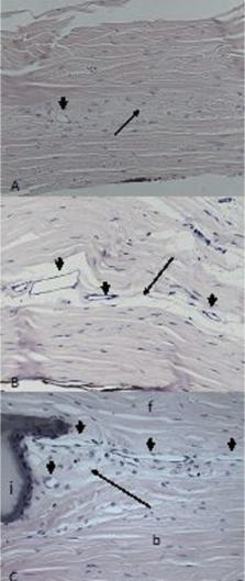
          The purpose of the study is an analysis of intrascleral drainage vessels formed in rabbits’ eyes after non-penetrating deep sclerectomy (NPDS) with absorbable and non-absorbable implants, and comparison to eyes in which surgery was performed without implanted material.

          Material/Methods

          NPDS was carried out in 12 rabbits, with implantation of non-absorbable methacrylic hydrogel (N=10 eyes) or absorbable cross-linked sodium hyaluronate (N=6 eyes), or without any implant (N=8 eyes). All the animals were euthanized 1 year after surgery. Twenty-one eyeballs were prepared for light microscopy and 3 were prepared for transmission electron microscope (TEM) analysis. Aqueous humour pathways were stained with ferritin in 6 eyeballs.

          Results

          By light microscopy, small vessels adjacent to the areas of scarring were the most common abnormality. Vessel density was significantly higher in operated sclera compared to normal, healthy tissue, regardless of the type of implant used. The average vessel densities were 2.18±1.48 vessels/mm 2 in non-implanted sclera, 2.34±1.69 vessels/mm 2 in eyes with absorbable implants, and 3.64±1.78 vessels/mm 2 in eyes with non-absorbable implants. Analysis of iron distribution in ferritin-injected eyes showed a positive reaction inside new aqueous draining vessels in all groups. TEM analysis showed that the ultrastructure of new vessels matched the features of the small veins.

          Conclusions

          Aqueous outflow after NPDS can be achieved through the newly formed network of small intrascleral veins. Use of non-absorbable implants significantly increases vessel density in the sclera adjacent to implanted material compared to eyes in which absorbable implants or no implants were used.

          Related collections

          Most cited references18

          • Record: found
          • Abstract: found
          • Article: not found

          Reducing capsular thickness and enhancing angiogenesis around implant drug release systems.

          Biological encapsulation and the foreign body reaction can impair the performance of implanted drug release devices. In this article, the classic definition of biocompatibility is questioned. Examples are presented of biomaterials showing unique healing behavior. A new paradigm for biomaterials healing is proposed in which non-specific protein adsorption is inhibited and matricellular proteins are controlled at the surfaces of implants.
            Bookmark
            • Record: found
            • Abstract: found
            • Article: not found

            Comparative study between deep sclerectomy with and without collagen implant: long term follow up.

            To identify the value of using collagen implant in deep sclerectomy. A prospective randomised trial of 104 eyes (104 patients) with medically uncontrolled primary and secondary open angle glaucoma. All patients had deep sclerectomy (DS), half of them with and the other half without a collagen implant (CI) sutured in the scleral bed. The main outcome measures were intraocular pressure (IOP), visual acuity, number of treatments preoperative and postoperative, and Nd:YAG goniopunctures. Mean follow up period was 44.5 (SD 21) months for the DS group and 43.9 (SD 14) months for the deep sclerectomy with a collagen implant (DSCI) group. The mean preoperative IOP was 23.3 (SD 7.2) mm Hg for the DS group and 25.6 (SD 4.9) mm Hg for the DSCI group. The mean IOP at the first postoperative day was 6.1 (SD 4.21) mm Hg for the DS group and 5.1 (SD 3.3) mm Hg for the DSCI group. At 48 months IOP was reduced by 40% (14 versus 23.3 mm Hg) for the DS group and by 50% (12.7 versus 25.6 mm Hg) for the DSCI group. Complete success rate, defined as IOP lower than 21 mm Hg without medication, was 34.6% (18/52 patients) at 48 months for the DS group, and 63.4% (33/52 patients) for the DSCI group. Qualified success rate; patients who achieved IOP below 21 mm Hg with or without medication, was 78.8% (41/52 patients) at 48 months and 94% (49/52 patients) for the DSCI group. The mean number of medications was reduced from 2.1 (SD 0.8) to 1.0 (SD 1) after DS, and was reduced from 2.2 (SD 0.7) to 0.4 (SD 0.6) in the DSCI group (p = 0.001) The use of a collagen implant in DS enhances the success rates and lowers the need for postoperative medication.
              Bookmark
              • Record: found
              • Abstract: found
              • Article: not found

              Deep sclerectomy in one eye vs deep sclerectomy with collagen implant in the contralateral eye of the same patient: long-term follow-up.

              To compare prospectively the results of deep sclerectomy (DS) vs deep sclerectomy with collagen implant (DSCI). Randomized prospective trial involving 26 eyes (13 patients) with medically uncontrolled primary and secondary open angle glaucoma. Collagen implant was randomly assigned to one eye of each patient. The mean follow-up period was 49.5 (SD 20) months for the DS-treated eyes, and 56.5 (SD 14) months for the DSCI-treated eyes (P=0.4). The mean preoperative intraocular pressure (IOP) was 24.1 (SD 7) mmHg for the DS-treated eyes, and 25.3 (SD 6) mmHg for the DSCI-treated eyes (P=0.5). The mean IOP at the first postoperative day was 6.4 (SD 3) mmHg for the DS-treated eyes, and 3.7 (SD 2) mmHg for the DSCI-treated eyes (P=0.05). The mean IOP at 12 months postoperative day was 15.4 (SD 3) mmHg for the DS group, and 10.4 (SD 4) mmHg for the DSCI-treated eyes (P=0.04), while at 48 months it was 16 (SD 3) mmHg for the DS group, and 10 (SD 4) mmHg for the DSCI-treated eyes (P=0.005). Complete success rate, defined as an IOP lower than 21 mmHg without medication, was 38% (5/13 patients) at 48 months for the DS-treated eyes, and 69% (9/13 patients) for the DSCI-treated eyes. Qualified success rate: patients who achieved IOP below 21 mmHg with or without medication, was 69% (9/13 patients) at 48 months and 100% (13/13 patients) for the DSCI group. The mean number of medications was reduced from 2.4 (SD 0.8) to 1.1 (SD 1) after DS, and was reduced from 2.2 (SD 0.7) to 0.4 (SD 0.6) in the DSCI group (P=0.001). For those eyes treated with DSCI, IOP was 3.21 mmHg lower than for those treated with DS (P<0.0001). The use of a collagen implant in DS seems to enhance the success rates, provides significantly lower IOP levels, and lowers the need for postoperative medications.
                Bookmark

                Author and article information

                Journal
                Med Sci Monit
                Med. Sci. Monit
                Medical Science Monitor
                Medical Science Monitor : International Medical Journal of Experimental and Clinical Research
                International Scientific Literature, Inc.
                1234-1010
                1643-3750
                2012
                01 October 2012
                : 18
                : 10
                : BR402-BR408
                Affiliations
                [1 ]Department of Ophthalmology, Collegium Medicum, Nicolaus Copernicus University, Bydgoszcz, Poland
                [2 ]Department of Pathology, Collegium Medicum, Nicolaus Copernicus University, Bydgoszcz, Poland
                [3 ]Department of Histology and Embryology, Collegium Medicum, Nicolaus Copernicus University, Bydgoszcz, Poland
                Author notes
                Jakub J. Kałużny, Department of Ophthalmology, Collegium Medicum, Nicolaus Copernicus University, Bydgoszcz, Poland, e-mail: kubeye@ 123456poczta.onet.pl
                [A]

                Study Design

                [B]

                Data Collection

                [C]

                Statistical Analysis

                [D]

                Data Interpretation

                [E]

                Manuscript Preparation

                [F]

                Literature Search

                [G]

                Funds Collection

                Article
                883480
                10.12659/MSM.883480
                3560554
                23018347
                6e4af031-035f-43d3-a3ae-6a09e3d3a515
                © Med Sci Monit, 2012

                This work is licensed under a Creative Commons Attribution-NonCommercial-NoDerivs 3.0 Unported License.

                History
                : 13 February 2012
                : 20 June 2012
                Categories
                Basic Research

                glaucoma,non-penetrating deep sclerectomy,aqueous outflow,intrascleral drainage vessels,intrascleral implant

                Comments

                Comment on this article