30
views
0
recommends
+1 Recommend
0 collections
    0
    shares
      • Record: found
      • Abstract: found
      • Article: found
      Is Open Access

      Comparative analysis of rodent and small mammal viromes to better understand the wildlife origin of emerging infectious diseases

      research-article

      Read this article at

      Bookmark
          There is no author summary for this article yet. Authors can add summaries to their articles on ScienceOpen to make them more accessible to a non-specialist audience.

          Abstract

          Background

          Rodents represent around 43% of all mammalian species, are widely distributed, and are the natural reservoirs of a diverse group of zoonotic viruses, including hantaviruses, Lassa viruses, and tick-borne encephalitis viruses. Thus, analyzing the viral diversity harbored by rodents could assist efforts to predict and reduce the risk of future emergence of zoonotic viral diseases.

          Results

          We used next-generation sequencing metagenomic analysis to survey for a range of mammalian viral families in rodents and other small animals of the orders Rodentia, Lagomorpha, and Soricomorpha in China. We sampled 3,055 small animals from 20 provinces and then outlined the spectra of mammalian viruses within these individuals and the basic ecological and genetic characteristics of novel rodent and shrew viruses among the viral spectra. Further analysis revealed that host taxonomy plays a primary role and geographical location plays a secondary role in determining viral diversity. Many viruses were reported for the first time with distinct evolutionary lineages, and viruses related to known human or animal pathogens were identified. Phylogram comparison between viruses and hosts indicated that host shifts commonly happened in many different species during viral evolutionary history.

          Conclusions

          These results expand our understanding of the viromes of rodents and insectivores in China and suggest that there is high diversity of viruses awaiting discovery in these species in Asia. These findings, combined with our previous bat virome data, greatly increase our knowledge of the viral community in wildlife in a densely populated country in an emerging disease hotspot.

          Electronic supplementary material

          The online version of this article (10.1186/s40168-018-0554-9) contains supplementary material, which is available to authorized users.

          Related collections

          Most cited references45

          • Record: found
          • Abstract: found
          • Article: not found

          The delayed rise of present-day mammals.

          Did the end-Cretaceous mass extinction event, by eliminating non-avian dinosaurs and most of the existing fauna, trigger the evolutionary radiation of present-day mammals? Here we construct, date and analyse a species-level phylogeny of nearly all extant Mammalia to bring a new perspective to this question. Our analyses of how extant lineages accumulated through time show that net per-lineage diversification rates barely changed across the Cretaceous/Tertiary boundary. Instead, these rates spiked significantly with the origins of the currently recognized placental superorders and orders approximately 93 million years ago, before falling and remaining low until accelerating again throughout the Eocene and Oligocene epochs. Our results show that the phylogenetic 'fuses' leading to the explosion of extant placental orders are not only very much longer than suspected previously, but also challenge the hypothesis that the end-Cretaceous mass extinction event had a major, direct influence on the diversification of today's mammals.
            Bookmark
            • Record: found
            • Abstract: found
            • Article: found
            Is Open Access

            Detection of Zoonotic Pathogens and Characterization of Novel Viruses Carried by Commensal Rattus norvegicus in New York City

            INTRODUCTION Zoonotic pathogens comprise a significant and increasing proportion of all new and emerging human infectious diseases (1, 2). Although zoonotic transmission is influenced by many factors, the frequency of contact between animal reservoirs and the human population appears to be a key element (3). Therefore, the risk of zoonotic transmission is increased by events that act to reduce the geographic or ecological separation between human and animal populations or increase the density and abundance of these populations where they coexist (2, 4). In this context, rapid and continuous urbanization constitutes a significant challenge to human health, as it creates irreversible changes to biodiversity that are driven by varied responses from animal species. In particular, species classified as urban exploiters and urban adapters may exist in unnaturally large and dense populations within urban environments and have above-average rates of contact with people (5 – 7). Of these, few species have been as successful at adapting to a peridomestic lifestyle as the Norway rat (Rattus norvegicus). In the urban environment, Norway rats closely cohabitate with humans—living inside buildings, feeding on refuse, and coming into contact with many aspects of the food supply (7 – 9). These characteristics, coupled with high levels of fecundity, growth rates, and population densities, suggest that urban Norway rats may be an important source of zoonotic pathogens (10, 11). Indeed, the Norway rat is a known reservoir of a range of human pathogens, including hantaviruses, Bartonella spp., and Leptospira interrogans; however, little is known about the microbial diversity present in urban rat populations or the risks they may pose to human health (12 – 16). As a first step toward understanding the zoonotic disease risk posed by rats in densely urban environments, we assessed the presence and prevalence of known and novel microbes in Norway rats in New York City (NYC). We took the unique approach of using both targeted molecular assays to detect known human pathogens and unbiased high-throughput sequencing (UHTS) to identify novel viruses related to agents of human disease. We quantified the tissue distribution of these novel viruses in the host using molecular methods and, in some cases, identified the site(s) of replication using strand-specific quantitative reverse transcription (RT)-PCR (ssqPCR). Unlike previous urban studies that have primarily relied on serological assays to assess the total prevalence of historic infection, our data provide a snapshot estimate of the current level of infection in the rat population, a parameter more closely related to the risk of zoonotic transmission (12, 17). Furthermore, as previous work has focused on rats found exclusively in outdoor locations, we concentrated our sampling within the built environment, where direct and indirect human-rodent contact is more likely to occur (18). RESULTS Sample collection. A total of 133 Norway rats were collected from five sites in NYC. Males (n = 72) were trapped slightly more often than females (n = 61), and juveniles were trapped more often than any other age category. Of the female rats, 43% were juveniles, 26% were subadults, and 31% were sexually mature adults, whereas 40% of the male rats were juveniles, 33% were subadults, and 26% were sexually mature. Targeted molecular analyses. Specific PCR-based assays were used to screen for the presence of 18 bacterial and 2 protozoan human pathogens (see Table S1 in the supplemental material). None of the samples tested was positive for Campylobacter coli, Listeria monocytogenes, Rickettsia spp., Toxoplasma gondii, Vibrio vulnificus, or Yersinia pestis, despite previous studies documenting most of these in multiple rodent species (15). All other bacterial and protozoan pathogens were detected in at least one animal (Table 1). Three bacterial pathogens were identified in more than 15% of animals: atypical enteropathogenic Escherichia coli (EPEC) was the most common (detected in 38% of rats), followed by Bartonella spp. (25% of rats) and Streptobacillus moniliformis (17% of rats) (Table 1). Phylogenetic analysis of a 327-nucleotide (nt) region of the gltA gene amplified from all infected animals revealed three distinct Bartonella species infecting NYC rats (Fig. S1). The most common of these was detected in 76% of Bartonella-positive animals and clustered within the Bartonella tribocorum group, which has previously been identified in multiple species of Rattus in Asia and North America. In addition, sequences 98% similar to those of Bartonella rochalimae were recovered from seven rats, and Bartonella elizabethae was identified in a single rat (Fig. S2). Infection with Bartonella was positively correlated with the age of the rat (P 90% similar at the nucleotide level to viruses known to infect Norway rats (e.g., Killham rat virus, rat astrovirus, and infectious diarrhea of infant rats [IDIR] agent [group B rotavirus]) and were not pursued further (Table 2). Viruses from an additional 13 families or genera that were 0.05). Only 10 rats were infected with more than two bacterial species, of which eight were female, and no rats were infected with more than four bacterial species (Table 4). In contrast, 53 rats were positive for more than two viral agents, and 13 of these carried more than five viruses (Table 4). As many as nine different viruses or four different bacterial species were identified in the same individual, with a maximum of 11 agents detected in a single rat. Patterns of coinfection between all agents were significantly nonrandom across the complete data set (C score, P = 0.0001); however, the only significantly positive association between any two bacteria occurred between Bartonella spp. and S. moniliformis (P = 0.005). Significantly positive associations were also observed between Bartonella spp. and multiple viruses, including NrKoV-1 and NrKoV-2 (P 180 g for females, >200 g for males) (65). The rats were necropsied, and the following tissues aseptically collected: brain, heart, kidney (83 rats only), liver, lung, inguinal lymph tissue, upper and lower intestine, salivary gland with associated lymph tissue, spleen, gonads (25 rats only), and urine or bladder (when <200 µl of urine was available). Oral and rectal swab samples were collected using sterile polyester swabs (Puritan Medical Products Company, Guilford, ME), and fecal pellets were collected when available. All samples were flash-frozen immediately following collection and stored at −80°C. All procedures described in this study were approved by the Institutional Animal Care and Use Committee at Columbia University (protocol number AC-AAAE6805). Targeted molecular analyses. DNA and RNA were extracted from each tissue and fecal sample using the AllPrep DNA/RNA minikit (Qiagen, Inc.) and from urine or serum using the QIAamp viral RNA minikit (Qiagen, Inc.). The extracted DNA was quantified and diluted to a working concentration of ≤400 ng/µl. Extracted RNA was quantified, and ≤5 µg used for cDNA synthesis with SuperScript III reverse transcriptase (Invitrogen) and random hexamers. Samples were tested by PCR for 10 bacterial, protozoan, and viral human pathogens previously associated with rodents using novel and previously published PCR assays, including Bartonella spp., L. interrogans, Rickettsia spp., S. moniliformis, Y. pestis, Cryptosporidium parvum, T. gondii, hepeviruses, hantaviruses (consensus assay), and SEOV (see Table S1 in the supplemental material). Each assay was performed using a subset of the sample types from each rat, selected to include known sites of replication or shedding (Table 1). Fecal samples were further analyzed for the presence of the following eight bacterial pathogens commonly associated with human gastrointestinal disease, using PCR-based assays: C. coli, Campylobacter jejuni, C. difficile, C. perfringens, L. monocytogenes, S. enterica, V. vulnificus, and Yersinia enterocolitica (see Table S1 in the supplemental material). PCR was also used to test for the presence of pathogenic E. coli, including enteroinvasive (EIEC, including Shigella), enterohemorrhagic (EHEC), enterotoxogenic (ETEC), enteroaggregative (EAEC), and enteropathogenic (EPEC) E. coli strains, using primers targeting virulence genes (Table S1) (66). In all cases, positive PCR products were confirmed by bidirectional dideoxy sequencing. Before Ro-SaV2 detection in intestinal samples was attempted, intestines were pretreated to remove fecal contamination by thorough washing with phosphate-buffered saline (PBS). To verify the absence of fecal material in the intestines, a PCR assay for cucumber green mottle mosaic virus (CGMMV) was performed on cDNA from paired fecal and intestinal samples from Ro-SaV2-infected animals. CGMMV was present in 10/11 Ro-SaV2-positive fecal samples and likely originated from the cucumber provided as a water source in the traps. However, all eight intestinal samples that were positive for Ro-SaV2 were negative for CGMVV, suggesting true intestinal infection by Ro-SaV2. UHTS. Serum samples and fecal pellets or rectal swab samples were also extracted, using a viral particle purification procedure, for UHTS. Briefly, each sample was successively passed through 0.45 µM and 0.22 µM sterile filters (Millipore) to remove bacterial and cellular debris and was treated with nucleases. Samples were lysed in NucliSENS buffer, extracted using the EasyMag platform (bioMérieux), and prepared for sequencing using the Ion Torrent Personal Genome Machine system, following the methods of Kapoor et al. (19). Sequencing was performed on pools of four to six samples, which were combined at the double-stranded DNA stage. Viral sequences were assembled using the Newbler or miraEST assemblers, and both contigs and unassembled reads were identified by similarity searches using BLASTn and BLASTx against the GenBank nonredundant nucleotide sequence database (67, 68). Viruses related to those known to cause disease in humans were selected for further study and verified by PCR on original (unpooled) sample material with primers derived from the UHTS sequence data. Confirmed positive results were followed by testing of the serum (n = 114) or fecal samples (n = 133) from remaining animals, and in some cases, subsequent screening of additional sample types from select positive animals (Table 3). One or more positive samples were chosen for further sequencing of phylogenetically relevant genes by overlapping PCR. The 5′ UTRs of NrPV, MPeV, and RPV were determined by rapid amplification of cDNA ends (RACE) using the SMARTer RACE cDNA amplification kit (Clontech). SEOV Baxter qPCR. Primers were designed to target a 121-nt region of the N gene (Baxter.qF, 5′ CATACCTCAGACGCACAC 3′; Baxter.qR, 5′ GGATCCATGTCATCACCG 3′; and Baxter. Probe, 5′-[6-FAM]CCTGGGGAAAGGAGGCAGTGGAT[TAMRA]-3′ [6-FAM, 6-carboxyfluorescein; TAMRA, 6-carboxytetramethylrhodamine]). For tissue samples, viral RNA copy numbers were normalized to the quantity of the reference gene encoding glyceraldehyde 3-phosphate dehydrogenase (GAPDH), whereas the viral RNA copy numbers in serum, oral, and rectal swab samples were reported per ml of serum or PBS wash, respectively (69). qPCR assays were run in duplicate on each sample, and the results were averaged. Samples with an average of ≤2 normalized copies were considered negative. ssqPCR. For ssqPCR, strand-specific synthetic standards were generated by transcribing positive- and negative-sense RNA in vitro from pCRII-TOPO dual promoter vectors (Life Technologies) containing 310 and 594 nt of the NS3 genes of NrHV-1 and NrHV-2, respectively. Positive- and negative-sense RNAs were synthesized from HindIII- or EcoRV-linearized plasmids by transcription from the T7 or SP6 RNA polymerase promoter. In vitro transcription was carried out for 2 h at 37°C using the RiboMax large-scale RNA production system (Promega) and 500 ng of linearized plasmid. Plasmid DNA was removed from the synthetic RNA transcripts by treatment with DNase I (Promega) for 30 min, followed by purification with the High Pure RNA purification kit (Roche). Purified RNA transcripts were analyzed on the Agilent 2100 Bioanalyzer, and RNA standards were prepared by serial dilution in human total RNA. cDNA from both strands was generated using strand-specific primers containing a tag sequence at the 5′ end (see Table S2 in the supplemental material) (70). The RNA was preheated at 70°C for 5 min with 10 pmol of specific primer and 1× reverse transcriptase buffer, followed by the addition of a preheated reaction mixture containing 1 mM MnCl2, 200 µM each deoxynucleoside triphosphate (dNTP), 40 U RNaseOUT, and 1 U Tth DNA polymerase (Promega). The reaction mixtures were incubated at 62°C for 2 min, followed by 65°C for 30 min. The cDNA was incubated with preheated 1× chelate buffer at 98°C for 30 min to inactivate the Tth reverse transcriptase before exonuclease I treatment to remove unincorporated RT primers (New England Biolabs). Reaction mixtures lacking RT primer were included to control for self-priming, the strand specificity of each primer was assessed by performing the RT step in the presence of the uncomplementary strand, and reaction mixtures lacking Tth DNA polymerase were included to control for plasmid DNA detection. ssqPCRs were performed using TaqMan universal master mix II with primers, probe, and 2 µl of cDNA under the following conditions: 50°C for 2 min, 95°C for 10 min, and then 40 cycles of 95°C for 15 s, 50°C for 20 s, and 72°C for 30 s (see Table S2 in the supplemental material). The specificity of the reaction was monitored by RT and amplification of serial dilutions of the uncomplementary strand. The sensitivities of the ssqPCR assays ranged from 0.35 × 10³ to 3.5 × 10³ RNA copies/reaction mixture volume, and nonstrand-specific amplification was not detected until 3.5 × 10⁷ viral RNA copies of the uncomplementary strand per reaction mixture volume were present (Table S3). Phylogenetic and sequence analyses. Nucleotide or predicted amino acid sequences were aligned with representative members of the relevant family or genus using MUSCLE in Geneious version 7 (Biomatters Ltd.) and manually adjusted. Maximum-likelihood (ML) and Bayesian Markov chain Monte Carlo (MCMC) phylogenetic trees were constructed for each alignment using RAxML version 8.0 and MrBayes version 3.2, respectively (71, 72). ML trees were inferred using the rapid-search algorithm, either the general time-reversible (GTR) plus gamma model of nucleotide substitution or the Whelan and Goldman (WAG) plus gamma model of amino acid substitution, and 500 bootstrap replicates. MCMC trees were inferred using the substitution models described above, a minimum of 10 million generations with sampling every 10,000 generations and terminated when the standard deviation of split frequencies reached <0.01. Phylogenetic analysis of Bartonella was performed by trimming the gltA gene sequences to a 327-nt region (nt positions 801 to 1127) commonly used for taxonomic classification and constructing a neighbor-joining tree using the Hasegawa, Kishino, and Yano (HKY) plus gamma model of nucleotide substitution (13, 73). Phylogenetic analysis of the flaviviruses was performed by first constructing a tree that included representative viruses across the family using a highly conserved region of the NS5B protein (aa 462 to 802 of tick-borne encephalitis virus; GenBank accession number NP_775511.1), followed by complete NS3 and NS5B amino acid phylogenies constructed separately for the Pestivirus and Hepacivirus/Pegivirus genera. These were rooted based on the relative positions of each genus in the family-level tree. To estimate the temporal and geographic origin of SEOV Baxter in NYC, phylogeographic analysis of the N gene was performed using the MCMC method available in the BEAST package (version 1.8.0) (74). All available full-length or nearly full-length SEOV N gene sequences with published sampling times were downloaded from GenBank, aligned as described above, and randomly subsampled five times to include a maximum of 10 sequences per country per year. The codon-structured SDR06 model of nucleotide substitution was used along with a relaxed, uncorrelated lognormal molecular clock and a constant population size coalescent prior (best-fit model, data not shown). Two independent MCMC chains were run for 100 million generations each, and convergence of all relevant parameters was assessed using Tracer version 1.5. The runs were combined after removing a 10% burn-in, and the maximum-clade-credibility (MCC) tree, including ancestral location-state reconstructions, was summarized. Transmembrane domain prediction was performed using TMHHM 2.0, putative N-glycosylation sites were predicted with NetNGlyc 1.0, and the presence of N-terminal signal peptides was predicted using SignalP 4.1, all of which were accessed through the ExPASy web portal (http://www.expasy.org). RNA secondary structures were predicted by MFOLD and through homology searching and structural alignment with bases conserved in other pestiviruses for NrPV and with parechoviruses, hunniviruses, and rosavirus for RPV (75). RNA structures were initially drawn using PseudoViewer, followed by manual editing (76). Statistical analyses. Potential associations between the presence of a microbial agent and the age and sex of the rat were explored using chi-square tests performed with SPSS version 21 (IBM, Armonk, NY), with associations considered significant at a level of α = 0.05. Tests for the overall effect of the age category or sex on the number of viruses carried by an individual were conducted using the Kruskal-Wallis test for age and the Wilcoxon test for sex. Patterns of pathogen cooccurrence within a single host were explored using the Fortran software program PAIRS version 1.1, which utilizes a Bayesian approach to detect nonrandom associations between pairs of taxa (77). The C score statistic was employed as a measure of pathogen cooccurrence (78). Nucleotide sequence accession numbers. The GenBank accession numbers for the agents sequenced in this study are KJ950830 to KJ951004. SUPPLEMENTAL MATERIAL Figure S1 Neighbor-joining tree of a 327-nt region of the gltA gene of Bartonella. The sequences derived from this study are indicated by a circle, and those recovered from Rattus norvegicus rats in Los Angeles, China, and Southeast Asia are given by squares and triangles, respectively. Download Figure S1, PDF file, 0.3 MB Figure S2 Predicted RNA secondary structure of the 5′ UTR of NrPV. Completely conserved nucleotides across all pestiviruses are colored in pink, and the conserved structural domains are indicated. Numbers indicate nucleotide positions from the 5′ end. Download Figure S2, PDF file, 0.5 MB Figure S3 (A) Predicted RNA secondary structure of the 5′ UTR of RPV. Completely conserved nucleotides across all parechovirus and hunnivirus 5′ UTR sequences are colored in pink, and the conserved structural domains are indicated. Numbers indicate nucleotide positions from the 5′ end. (B) ML tree of the complete VP1 gene of RPV and feline, bat, and canine picornaviruses. Selected representatives of the Enterovirus and Sapelovirus genera are also shown. When the BSP and BPP values are both ≥70%, the nodal support value is given beneath associated nodes in the format BSP/BPP. Download Figure S3, PDF file, 0.8 MB Figure S4 Unrooted ML tree of the complete VP1 gene of Manhattan parechovirus (MPeV) (indicated by a red branch) and representative members of the Parechovirus genus. When the BSP and BPP values are both ≥70%, the nodal support is shown beneath the associated node in the format BSP/BPP. HPeV, human parechovirus. Download Figure S4, PDF file, 0.3 MB Table S1 Primers used for targeted molecular analysis in this study. Table S1, DOCX file, 0.1 MB. Table S2 Primer and probe sequences used in the strand-specific reverse transcription and quantitative PCR assays of NrHV-1 and NrHV-2. Table S2, DOCX file, 0.1 MB. Table S3 Sensitivities and specificities of the strand-specific qPCR assays for NrHV-1 and NrHV-2. Table S3, DOCX file, 0.04 MB.
              Bookmark
              • Record: found
              • Abstract: found
              • Article: found
              Is Open Access

              Genetic Detection and Characterization of Lujo Virus, a New Hemorrhagic Fever–Associated Arenavirus from Southern Africa

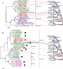
              Introduction Members of the genus Arenavirus, comprising currently 22 recognized species (http://www.ictvonline.org/virusTaxonomy.asp?version=2008), are divided into two complexes based on serologic, genetic, and geographic relationships [1],[2]: the New World (NW) or Tacaribe complex, and the Old World (OW) or Lassa-Lymphocytic choriomeningitis complex that includes the ubiquitous arenavirus type-species Lymphocytic choriomeningitis virus (LCMV; [3]). The RNA genome of arenaviruses is bi-segmented, comprising a large (L) and a small (S) segment that each codes for two proteins in ambisense coding strategy [4],[5]. Despite this coding strategy, the Arenaviridae are classified together with the families Orthomyxoviridae and Bunyaviridae as segmented single-strand, negative sense RNA viruses. The South American hemorrhagic fever viruses Junin (JUNV; [6],[7]), Machupo (MACV; [8]), Guanarito (GTOV; [9]) and Sabia virus (SABV, [10]), and the African Lassa virus (LASV [11]), are restricted to biosafety level 4 (BSL-4) containment due to their associated aerosol infectivity and rapid onset of severe disease. With the possible exception of NW Tacaribe virus (TCRV; [12]), which has been isolated from bats (Artibeus spp.), individual arenavirus species are commonly transmitted by specific rodent species wherein the capacity for persistent infection without overt disease suggests long evolutionary adaptation between the agent and its host [1], [13]–[16]. Whereas NW arenaviruses are associated with rodents in the Sigmodontinae subfamily of the family Cricetidae, OW arenaviruses are associated with rodents in the Murinae subfamily of the family Muridae. Humans are most frequently infected through contact with infected rodent excreta, commonly via inhalation of dust or aerosolized virus-containing materials, or ingestion of contaminated foods [13]; however, transmission may also occur by inoculation with infected body fluids and tissue transplantation [17]–[19]. LCMV, which is spread by the ubiquitous Mus musculus as host species and hence found world-wide, causes symptoms in humans that range from asymptomatic infection or mild febrile illness to meningitis and encephalitis [13]. LCMV infection is only rarely fatal in immunocompetent adults; however, infection during pregnancy bears serious risks for mother and child and frequently results in congenital abnormalities. The African LASV, which has its reservoir in rodent species of the Mastomys genus, causes an estimated 100,000–500,000 human infections per year in West African countries (Figure 1). Although Lassa fever is typically sub-clinical or associated with mild febrile illness, up to 20% of cases may have severe systemic disease culminating in fatal outcome [20],[21]. Three other African arenaviruses are not known to cause human disease: Ippy virus (IPPYV; [22],[23]), isolated from Arvicanthis spp. and Mobala virus (MOBV; [24]) isolated from Praomys spp. in the Central African Republic (CAR); and Mopeia virus (MOPV) that like LASV is associated with members of the genus Mastomys, and was reported from Mozambique [25] and Zimbabwe [26], although antibody studies suggest that MOPV and LASV may also circulate in CAR [27] where the geographies of these viruses appear to overlap (Figure 1). Up to present, there have been no published reports of severe human disease associated with arenaviruses isolated from southern Africa. 10.1371/journal.ppat.1000455.g001 Figure 1 Geographic distribution of African arenaviruses. MOBV, MOPV, and IPPYV (blue) have not been implicated in human disease; LASV (red) can cause hemorrhagic fever. The origin of the LUJV index and secondary and tertiary cases linked in the 2008 outbreak are indicated in gold. In September 2008 an outbreak of unexplained hemorrhagic fever was reported in South Africa [28]. The index patient was airlifted in critical condition from Zambia on September 12 to a clinic in Sandton, South Africa, after infection from an unidentified source. Secondary infections were recognized in a paramedic (case 2) who attended the index case during air transfer from Zambia, in a nurse (case 3) who attended the index case in the intensive care unit in South Africa, and in a member of the hospital staff (case 4) who cleaned the room after the index case died on September 14. One case of tertiary infection was recorded in a nurse (case 5) who attended case 2 after his transfer from Zambia to Sandton on September 26, one day before barrier nursing was implemented. The course of disease in cases 1 through 4 was fatal; case 5 received ribavirin treatment and recovered. A detailed description of clinical and epidemiologic data, as well as immunohistological and PCR analyses that indicated the presence of an arenavirus, are reported in a parallel communication (Paweska et al., Emerg. Inf. Dis., submitted). Here we report detailed genetic analysis of this novel arenavirus. Results/Discussion Rapid identification of a novel pathogen through unbiased pyrosequencing RNA extracts from two post-mortem liver biopsies (cases 2 and 3) and one serum sample (case 2) were independently submitted for unbiased high-throughput pyrosequencing. The libraries yielded between 87,500 and 106,500 sequence reads. Alignment of unique singleton and assembled contiguous sequences to the GenBank database (http://www.ncbi.nlm.nih.gov/Genbank) using the Basic Local Alignment Search Tool (blastn and blastx; [29]) indicated coverage of approximately 5.6 kilobases (kb) of sequence distributed along arenavirus genome scaffolds: 2 kb of S segment sequence in two fragments, and 3.6 kb of L segment sequence in 7 fragments (Figure 2). The majority of arenavirus sequences were obtained from serum rather than tissue, potentially reflecting lower levels of competing cellular RNA in random amplification reactions. 10.1371/journal.ppat.1000455.g002 Figure 2 LUJV genome organization and potential secondary structure of intergenic regions. Open reading frames (ORF) for the glycoprotein precursor GPC, the nucleoprotein NP, the matrix protein analog Z, and the polymerase L, and their orientation are indicated (A); blue bars represent sequences obtained by pyrosequencing from clinical samples. Secondary structure predictions of intergenic regions (IR) for S (B, C) and L segment sequence (D, E) in genomic (B, D) and antigenomic orientation (C, E) were analyzed by mfold; shading indicates the respective termination codon (opal, position 1), and its reverse-complement, respectively. Full genome characterization of a newly identified arenavirus Sequence gaps between the aligned fragments were rapidly filled by specific PCR amplification with primers designed on the pyrosequence data at both, CU and CDC. Terminal sequences were added by PCR using a universal arenavirus primer, targeting the conserved viral termini (5′-CGC ACM GDG GAT CCT AGG C, modified from [30]) combined with 4 specific primers positioned near the ends of the 2 genome segments. Overlapping primer sets based on the draft genome were synthesized to facilitate sequence validation by conventional dideoxy sequencing. The accumulated data revealed a classical arenavirus genome structure with a bi-segmented genome encoding in an ambisense strategy two open reading frames (ORF) separated by an intergenic stem-loop region on each segment (Figure 2) (GenBank Accession numbers FJ952384 and FJ952385). Our data represent genome sequences directly obtained from liver biopsy and serum (case 2), and from cell culture isolates obtained from blood at CDC (case 1 and 2), and from liver biopsies at NICD (case 2 and 3). No sequence differences were uncovered between virus detected in primary clinical material and virus isolated in cell culture at the two facilities. In addition, no changes were detected between each of the viruses derived from these first three cases. This lack of sequence variation is consistent with the epidemiologic data, indicating an initial natural exposure of the index case, followed by a chain of nosocomial transmission among subsequent cases. Lujo virus (LUJV) is a novel arenavirus Phylogenetic trees constructed from full L or S segment nucleotide sequence show LUJV branching off the root of the OW arenaviruses, and suggest it represents a highly novel genetic lineage, very distinct from previously characterized virus species and clearly separate from the LCMV lineage (Figure 3A and 3B). No evidence of genome segment reassortment is found, given the identical placement of LUJV relative to the other OW arenaviruses based on S and L segment nucleotide sequences. In addition, phylogenetic analysis of each of the individual ORFs reveals similar phylogenetic tree topologies. A phylogenetic tree constructed from deduced L-polymerase amino acid (aa) sequence also shows LUJV near the root of the OW arenaviruses, distinct from characterized species, and separate from the LCMV branch (Figure 3C). A distant relationship to OW arenaviruses may also be inferred from the analysis of Z protein sequence (Figure S1). The NP gene sequence of LUJV differs from other arenaviruses from 36% (IPPYV) to 43% (TAMV) at the nucleotide level, and from 41% (MOBV/LASV) to 55% (TAMV) at the aa level (Table S1). This degree of divergence is considerably higher than both, proposed cut-off values within ( 21.5%) OW arenavirus species [31],[32], and indicates a unique phylogenitic position for LUJV (Figure 3D). Historically, phylogenetic assignments of arenaviruses have been based on portions of the NP gene [1],[33], because this is the region for which most sequences are known. However, as more genomic sequences have become available, analyses of full-length GPC sequence have revealed evidence of possible relationships between OW and NW arenaviruses not revealed by NP sequence alone [34]. Because G1 sequences are difficult to align some have pursued phylogenetic analyses by combining the GPC signal peptide and the G2 sequence for phylogenetic analysis [16]. We included in our analysis the chimeric signal/G2 sequence (Figure 3E) as well as the receptor binding G1 portion (Figure 3F); both analyses highlighted the novelty of LUJV, showing an almost similar distance from OW as from NW viruses. 10.1371/journal.ppat.1000455.g003 Figure 3 Phylogenetic analyses of LUJV. Phylogenetic relationships of LUJV were inferred based on full L (A) and S segment nucleotide sequence (B), as well as on deduced amino acid sequences of L (C), NP (D), Signal/G2 (E) and G1 (F) ORF's. Phylogenies were reconstructed by neighbor-joining analysis applying a Jukes-Cantor model; the scale bar indicates substitutions per site; robust boostrap support for the positioning of LUJV was obtained in all cases (>98% of 1000 pseudoreplicates). GenBank Accession numbers for reference sequences are: ALLV CLHP2472 (AY216502, AY012687); AMAV BeAn70563 (AF512834); BCNV AVA0070039 (AY924390, AY922491), A0060209 (AY216503); CATV AVA0400135 (DQ865244), AVA0400212 (DQ865245); CHPV 810419 (EU, 260464, EU260463); CPXV BeAn119303 (AY216519, AF512832); DANV 0710-2678 (EU136039, EU136038); FLEV BeAn293022 (EU627611, AF512831); GTOV INH-95551 (AY358024, AF485258), CVH-960101 (AY497548); IPPYV DakAnB188d (DQ328878, DQ328877); JUNV MC2 (AY216507, D10072), XJ13 (AY358022, AY358023), CbalV4454 (DQ272266); LASV LP (AF181853), 803213 (AF181854), Weller (AY628206), AV (AY179171, AF246121), Z148 (AY628204, AY628205), Josiah (U73034, J043204), NL (AY179172, AY179173); LATV MARU10924 (EU627612, AF485259); LCMV Armstrong (AY847351), ARM53b (M20869), WE (AF004519, M22138), Marseille12 (DQ286932, DQ286931), M1 (AB261991); MACV Carvallo (AY619642, AY619643), Chicava (AY624354, AY624355), Mallele (AY619644, AY619645), MARU222688 (AY922407), 9530537 (AY571959); MOBV ACAR3080MRC5P2 (DQ328876, AY342390); MOPV AN20410 (AY772169, AY772170), Mozambique (DQ328875, DQ328874); NAAV AVD1240007 (EU123329); OLVV 3229-1 (AY216514, U34248); PARV 12056 (EU627613, AF485261); PICV (K02734), MunchiqueCoAn4763 (EF529745, EF529744), AN3739 (AF427517); PIRV VAV-488 (AY216505, AF277659); SABV SPH114202 (AY358026, U41071); SKTV AVD1000090 (EU123328); TAMV W10777 (EU627614, AF512828); TCRV (J04340, M20304); WWAV AV9310135 (AY924395, AF228063). Protein motifs potentially relevant to LUJV biology Canonical polymerase domains pre-A, A, B, C, D, and E [35]–[37] are well conserved in the L ORF of LUJV (256 kDa, pI = 6.4; Figure 4). The Z ORF (10.5 kDa, pI = 9.3) contains two late domain motifs like LASV; however, in place of the PTAP motif found in LASV, that mediates recognition of the tumor susceptibility gene 101, Tsg101 [38], involved in vacuolar protein sorting [39],[40], LUJV has a unique Y77REL motif that matches the YXXL motif of the retrovirus equine infectious anemia virus [41], which interacts with the clathrin adaptor protein 2 (AP2) complex [42]. A Tsg101-interacting motif, P90SAP, is found in LUJV in position of the second late domain of LASV, PPPY, which acts as a Nedd4-like ubiquitin ligase recognition motif [43]. The RING motif, containing conserved residue W44 [44], and the conserved myristoylation site G2 are present [45]–[47] (Figure 4). The NP of LUJV (63.1 kDa, pI = 9.0) contains described aa motifs that resemble mostly OW arenaviruses [48], including a cytotoxic T-lymphocyte (CTL) epitope reported in LCMV (GVYMGNL; [49]), corresponding to G122VYRGNL in LUJV, and a potential antigenic site reported in the N-terminal portion of LASV NP (RKSKRND; [50]), corresponding to R55KDKRND in LUJV (Figure 4). 10.1371/journal.ppat.1000455.g004 Figure 4 Schematic of conserved protein motifs. Conservation of LUJV amino acid motifs with respect to all other (green highlight), to OW (yellow highlight), or to NW (blue highlight) arenaviruses is indicated; grey highlight indicates features unique to LUJV. Polymerase motifs pre-A (L1142), A (N1209), B (M1313), C (L1345), D (Q1386), and E (C1398) are indicated for the L ORF; potential myristoylation site G2, the RING motif H34/C76, and potential late domains YXXL an PSAP are indicated for the Z ORF; and myristoylation site G2, posttranslational processing sites for signalase (S59/S60) and S1P cleavage (RKLM221), CTL epitope (I32), zinc finger motif P415/G440, as well as conserved cysteine residues and glycosylations sites (Y) are indicated for GPC. * late domain absent in NW viruses and DANV; † PSAP or PTAP in NW viruses, except in PIRV and TCRV (OW viruses: PPPY); # G in all viruses except LCMV ( = A); ‡ D in NW clade A only; § conserved with respect to OW, and NW clade A and C; HD, hydrophobic domain; TM, transmembrane anchor. The GPC precursor (52.3 kDa, pI = 9.0) is cotranslationally cleaved into the long, stable signal peptide and the mature glycoproteins G1 and G2 [51]–[54]. Based on analogy to LASV [55] and LCMV [56], signalase would be predicted to cleave between D58 and S59 in LUJV. However, aspartate and arginine residues in the −1 and −3 positions, respectively, violate the (−3,−1)-rule [57]; thus, cleavage may occur between S59 and S60 as predicted by the SignalP algorithm. The putative 59 aa signal peptide of LUJV displays a conserved G2, implicated in myristoylation in JUNV [58], however, it is followed in LUJV by a non-standard valine residue in position +4, resembling non-standard glycine residues found in Oliveros virus (OLVV [59]) and Latino virus (LATV; http://www2.ncid.cdc.gov/arbocat/catalog-listing.asp?VirusID=263&SI=1). Conservation is also observed for aa residues P12 (except Amapari virus; AMAV [60]), E17 [61](except Pirital virus; PIRV [62]), and N20 in hydrophobic domain 1, as well as I32KGVFNLYK40SG, identified as a CTL epitope in LCMV WE (I32KAVYNFATCG; [63]) (Figure 4). Analogous to other arenaviruses, SKI-1/S1P cleavage C-terminal of RKLM221 is predicted to separate mature G1 (162 aa, 18.9 kDa, pI = 6.4) from G2 (233 aa, 26.8 kDa, pI = 9.5) [52],[53],[64]. G2 appears overall well conserved, including the strictly conserved cysteine residues: 6 in the luminal domain, and 3 in the cytoplasmic tail that are included in a conserved zinc finger motif reported in JUNV [65] (Figure 4). G2 contains 6 potential glycosylation sites, including 2 strictly conserved sites, 2 semi-conserved sites N335 (absent in LCMVs and Dandenong virus; DANV [19]) and N352 (absent in LATV), and 2 unique sites in the predicted cytoplasmic tail (Figure 4). G1 is poorly conserved among arenaviruses [16], and G1 of LUJV is no exception, being highly divergent from the G1 of the other arenaviruses, and shorter than that of other arenaviruses. LUJV G1 contains 6 potential glycosylation sites in positions comparable to other arenaviruses, including a conserved site N93HS (Figure 4), which is shifted by one aa in a motif that otherwise aligns well with OW arenaviruses and NW arenavirus clade A and C viruses. There is no discernable homology to other arenavirus G1 sequences that would point to usage of one of the two identified arenavirus receptors; Alpha-dystroglycan (α-DG) [66] that binds OW arenaviruses LASV and LCMV, and NW clade C viruses OLVV and LATV [67], or transferrin receptor 1 (TfR1) that binds pathogenic NW arenaviruses JUNV, MACV, GTOV, and SABV [68] (Figure S2). In summary, our analysis of the LUJV genome shows a novel virus that is only distantly related to known arenaviruses. Sequence divergence is evident across the whole genome, but is most pronounced in the G1 protein encoded by the S segment, a region implicated in receptor interactions. Reassortment of S and L segments leading to changes in pathogenicity has been described in cultured cells infected with different LCMV strains [69], and between pathogenic LASV and nonpathogenic MOPV [70]. We find no evidence to support reassortment of the LUJV L or S genome segment (Figure 3A and 3B). Recombination of glycoprotein sequence has been recognized in NW arenaviruses [14], [16], [33], [34], [71]–[73], resulting in the division of the complex into four sublineages: lineages A, B, C, and an A/recombinant lineage that forms a branch of lineage A when NP and L sequence is considered (see Figure 3C and 3D), but forms an independent branch in between lineages B and C when glycoprotein sequence is considered (see Figure 3D). While recombination cannot be excluded in case of LUJV, our review of existing databases reveals no candidate donor for the divergent GPC sequence. To our knowledge is LUJV the first hemorrhagic fever-associated arenavirus from Africa identified in the past 3 decades. It is also the first such virus originating south of the equator (Figure 1). The International Committee on the Taxonomy of Viruses (ICTV) defines species within the Arenavirus genus based on association with a specific host, geographic distribution, potential to cause human disease, antigenic cross reactivity, and protein sequence similarity to other species. By these criteria, given the novelty of its presence in southern Africa, capacity to cause hemorrhagic fever, and its genetic distinction, LUJV appears to be a new species. Materials and Methods Sequencing Clinical specimens were inactivated in TRIzol (liver tissue, 100 mg) or TRIzol LS (serum, 250 µl) reagent (Invitrogen, Carlsbad, CA, USA) prior to removal from BSL-4 containment. Total RNA extracts were treated with DNase I (DNA-free, Ambion, Austin, TX, USA) and cDNA generated by using the Superscript II system (Invitrogen) and 100–500 ng RNA for reverse transcription primed with random octamers that were linked to an arbitrary, defined 17-mer primer sequence [74]. The resulting cDNA was treated with RNase H and then randomly amplified by the polymerase chain reaction (PCR; [75]); applying a 9∶1 mixture of a primer corresponding to the defined 17-mer sequence, and the random octamer-linked 17-mer primer, respectively [74]. Products >70 base pairs (bp) were selected by column purification (MinElute, Qiagen, Hilden, Germany) and ligated to specific linkers for sequencing on the 454 Genome Sequencer FLX (454 Life Sciences, Branford, CT, USA) without fragmentation of the cDNA [19],[76],[77]). Removal of primer sequences, redundancy filtering, and sequence assembly were performed with software programs accessible through the analysis applications at the GreenePortal website (http://156.145.84.111/Tools). Conventional PCRs at CU were performed with HotStar polymerase (Qiagen) according to manufacturer's protocols on PTC-200 thermocyclers (Bio-Rad, Hercules, CA, USA): an enzyme activation step of 5 min at 95°C was followed by 45 cycles of denaturation at 95°C for 1 min, annealing at 55°C for 1 min, and extension at 72°C for 1 to 3 min depending on the expected amplicon size. A two-step RT-PCR protocol was also followed at CDC using Invitrogen's Thermoscript RT at 60 degrees for 30 min followed by RNase H treatment for 20 min. cDNA was amplified using Phusion enzyme with GC Buffer (Finnzymes, Espoo, Finland) and 3% DMSO with an activation step at 98°C for 30 sec, followed by the cycling conditions of 98°C for 10 sec, 58°C for 20 sec, and 72°C for 1 min for 35 cycles and a 5 min extension at 72°C. Specific primer sequences are available upon request. Amplification products were run on 1% agarose gels, purified (MinElute, Qiagen), and directly sequenced in both directions with ABI PRISM Big Dye Terminator 1.1 Cycle Sequencing kits on ABI PRISM 3700 DNA Analyzers (Perkin-Elmer Applied Biosystems, Foster City, CA). Sequence analyses Programs of the Wisconsin GCG Package (Accelrys, San Diego, CA, USA) were used for sequence assembly and analysis; percent sequence difference was calculated based on Needleman-Wunsch alignments (gap open/extension penalties 15/6.6 for nucleotide and 10/0.1 for aa alignments; EMBOSS [78]), using a Perl script to iterate the process for all versus all comparison. Secondary RNA structure predictions were performed with the web-based version of mfold (http://mfold.bioinfo.rpi.edu); data were exported as .ct files and layout and annotation was done with CLC RNA Workbench (CLC bio, Århus, Denmark). Protein topology and targeting predictions were generated by employing SignalP, and NetNGlyc, TMHMM (http://www.cbs.dtu.dk/services), the web-based version of TopPred (http://mobyle.pasteur.fr/cgi-bin/portal.py?form=toppred), and Phobius (http://phobius.sbc.su.se/). Phylogenetic analyses were performed using MEGA software [79]. Supporting Information Figure S1 Phylogenetic tree based on deduced Z amino acid sequence. In contrast to phylogenetic trees obtained with the other ORFs (Figure 2), poor bootstrap support (43% of 1,000 pseudoreplicates) for the branching of LUJV off the LCMV clade was obtained with Z ORF sequence. For GenBank accession numbers see Figure 2. (0.44 MB TIF) Click here for additional data file. Figure S2 Pairwise sliding-window distance analysis of GPC sequence. LUJV and members of the OW (LASV, MOPV, IPPYV, LCMV, DANV) and NW (GTOV, CPXV, BNCV, PIRV, OLVV, SABV, MACV) arenavirus complex were compared using LASV NL (A) or GTOV CVH (B) as query (10 aa step; 80 aa window). (4.21 MB TIF) Click here for additional data file. Table S1 Pairwise nucleotide and amino acid differences between LUJV and other OW and NW arenaviruses. * NAAV, North American arenavirus. † Values <30% (amino acid) or <33% (nucleotide) are highlighted in green. (0.20 MB DOC) Click here for additional data file.
                Bookmark

                Author and article information

                Contributors
                daszak@ecohealthalliance.org
                wangjw28@163.com
                liuqiyong@icdc.cn
                zdsys@vip.sina.com
                Journal
                Microbiome
                Microbiome
                Microbiome
                BioMed Central (London )
                2049-2618
                3 October 2018
                3 October 2018
                2018
                : 6
                : 178
                Affiliations
                [1 ]ISNI 0000 0001 0662 3178, GRID grid.12527.33, MOH Key Laboratory of Systems Biology of Pathogens, Institute of Pathogen Biology, , Chinese Academy of Medical Sciences & Peking Union Medical College, ; Beijing, People’s Republic of China
                [2 ]ISNI 0000 0004 1759 700X, GRID grid.13402.34, Collaborative Innovation Center for Diagnosis and Treatment of Infectious Diseases, ; Hangzhou, People’s Republic of China
                [3 ]ISNI 0000 0000 8803 2373, GRID grid.198530.6, State Key Laboratory for Infectious Diseases Prevention and Control, National Institute for Communicable Disease Control and Prevention, , Chinese Center for Disease Control and Prevention, ; Beijing, People’s Republic of China
                [4 ]ISNI 0000 0004 0409 4702, GRID grid.420826.a, EcoHealth Alliance, ; New York, NY USA
                [5 ]ISNI 0000 0004 1758 1139, GRID grid.464500.3, Yunnan Institute of Parasitic Diseases, ; Puer, People’s Republic of China
                Article
                554
                10.1186/s40168-018-0554-9
                6171170
                30285857
                6d614ea1-d14b-4739-924f-f63b1823e90b
                © The Author(s). 2018

                Open AccessThis article is distributed under the terms of the Creative Commons Attribution 4.0 International License ( http://creativecommons.org/licenses/by/4.0/), which permits unrestricted use, distribution, and reproduction in any medium, provided you give appropriate credit to the original author(s) and the source, provide a link to the Creative Commons license, and indicate if changes were made. The Creative Commons Public Domain Dedication waiver ( http://creativecommons.org/publicdomain/zero/1.0/) applies to the data made available in this article, unless otherwise stated.

                History
                : 29 January 2018
                : 5 September 2018
                Funding
                Funded by: CAMS Innovation Fund for Medical Sciences
                Award ID: 2016-I2M-1-014
                Award Recipient :
                Funded by: FundRef http://dx.doi.org/10.13039/501100001809, National Natural Science Foundation of China;
                Award ID: 81772228
                Award ID: 81501773
                Award Recipient :
                Funded by: FundRef http://dx.doi.org/10.13039/100000060, National Institute of Allergy and Infectious Diseases;
                Award ID: R01AI110964
                Award Recipient :
                Funded by: Non-profit Central Institute Fund of Chinese Academy of Medical Sciences
                Award ID: 2017PT31013
                Award Recipient :
                Categories
                Research
                Custom metadata
                © The Author(s) 2018

                rodents,small mammals,virome,viral evolution,emerging infectious diseases

                Comments

                Comment on this article