14
views
0
recommends
+1 Recommend
1 collections
    0
    shares
      • Record: found
      • Abstract: found
      • Article: found
      Is Open Access

      An inhibitory gate for state transition in cortex

      research-article

      Read this article at

      Bookmark
          There is no author summary for this article yet. Authors can add summaries to their articles on ScienceOpen to make them more accessible to a non-specialist audience.

          Abstract

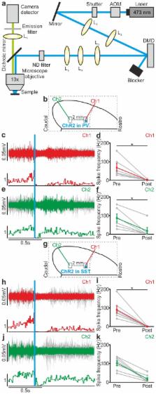
          Large scale transitions between active (up) and silent (down) states during quiet wakefulness or NREM sleep regulate fundamental cortical functions and are known to involve both excitatory and inhibitory cells. However, if and how inhibition regulates these activity transitions is unclear. Using fluorescence-targeted electrophysiological recording and cell-specific optogenetic manipulation in both anesthetized and non-anesthetized mice, we found that two major classes of interneurons, the parvalbumin and the somatostatin positive cells, tightly control both up-to-down and down-to-up state transitions. Inhibitory regulation of state transition was observed under both natural and optogenetically-evoked conditions. Moreover, perturbative optogenetic experiments revealed that the inhibitory control of state transition was interneuron-type specific. Finally, local manipulation of small ensembles of interneurons affected cortical populations millimetres away from the modulated region. Together, these results demonstrate that inhibition potently gates transitions between cortical activity states, and reveal the cellular mechanisms by which local inhibitory microcircuits regulate state transitions at the mesoscale.

          DOI: http://dx.doi.org/10.7554/eLife.26177.001

          Related collections

          Most cited references60

          • Record: found
          • Abstract: found
          • Article: not found

          High-Performance Genetically Targetable Optical Neural Silencing via Light-Driven Proton Pumps

          The ability to silence the activity of genetically specified neurons in a temporally precise fashion would open up the ability to investigate the causal role of specific cell classes in neural computations, behaviors, and pathologies. Here we show that members of the class of light-driven outward proton pumps can mediate very powerful, safe, multiple-color silencing of neural activity. The gene archaerhodopsin-31 (Arch) from Halorubrum sodomense enables near-100% silencing of neurons in the awake brain when virally expressed in mouse cortex and illuminated with yellow light. Arch mediates currents of several hundred picoamps at low light powers, and supports neural silencing currents approaching 900 pA at light powers easily achievable in vivo. In addition, Arch spontaneously recovers from light-dependent inactivation, unlike light-driven chloride pumps that enter long-lasting inactive states in response to light. These properties of Arch are appropriate to mediate the optical silencing of significant brain volumes over behaviourally-relevant timescales. Arch function in neurons is well tolerated because pH excursions created by Arch illumination are minimized by self-limiting mechanisms to levels comparable to those mediated by channelrhodopsins2,3 or natural spike firing. To highlight how proton pump ecological and genomic diversity may support new innovation, we show that the blue-green light-drivable proton pump from the fungus Leptosphaeria maculans 4 (Mac) can, when expressed in neurons, enable neural silencing by blue light, thus enabling alongside other developed reagents the potential for independent silencing of two neural populations by blue vs. red light. Light-driven proton pumps thus represent a high-performance and extremely versatile class of “optogenetic” voltage and ion modulator, which will broadly empower new neuroscientific, biological, neurological, and psychiatric investigations.
            Bookmark
            • Record: found
            • Abstract: found
            • Article: not found

            Prefrontal phase locking to hippocampal theta oscillations.

            The interactions between cortical and hippocampal circuits are critical for memory formation, yet their basic organization at the neuronal network level is not well understood. Here, we demonstrate that a significant portion of neurons in the medial prefrontal cortex of freely behaving rats are phase locked to the hippocampal theta rhythm. In addition, we show that prefrontal neurons phase lock best to theta oscillations delayed by approximately 50 ms and confirm this hippocampo-prefrontal directionality and timing at the level of correlations between single cells. Finally, we find that phase locking of prefrontal cells is predicted by the presence of significant correlations with hippocampal cells at positive delays up to 150 ms. The theta-entrained activity across cortico-hippocampal circuits described here may be important for gating information flow and guiding the plastic changes that are believed to underlie the storage of information across these networks.
              Bookmark
              • Record: found
              • Abstract: found
              • Article: not found

              Natural waking and sleep states: a view from inside neocortical neurons.

              In this first intracellular study of neocortical activities during waking and sleep states, we hypothesized that synaptic activities during natural states of vigilance have a decisive impact on the observed electrophysiological properties of neurons that were previously studied under anesthesia or in brain slices. We investigated the incidence of different firing patterns in neocortical neurons of awake cats, the relation between membrane potential fluctuations and firing rates, and the input resistance during all states of vigilance. In awake animals, the neurons displaying fast-spiking firing patterns were more numerous, whereas the incidence of neurons with intrinsically bursting patterns was much lower than in our previous experiments conducted on the intact-cortex or isolated cortical slabs of anesthetized cats. Although cortical neurons displayed prolonged hyperpolarizing phases during slow-wave sleep, the firing rates during the depolarizing phases of the slow sleep oscillation was as high during these epochs as during waking and rapid-eye-movement sleep. Maximum firing rates, exceeding those of regular-spiking neurons, were reached by conventional fast-spiking neurons during both waking and sleep states, and by fast-rhythmic-bursting neurons during waking. The input resistance was more stable and it increased during quiet wakefulness, compared with sleep states. As waking is associated with high synaptic activity, we explain this result by a higher release of activating neuromodulators, which produce an increase in the input resistance of cortical neurons. In view of the high firing rates in the functionally disconnected state of slow-wave sleep, we suggest that neocortical neurons are engaged in processing internally generated signals.
                Bookmark

                Author and article information

                Contributors
                Role: Reviewing editor
                Journal
                eLife
                Elife
                eLife
                eLife
                eLife
                eLife Sciences Publications, Ltd
                2050-084X
                16 May 2017
                2017
                : 6
                : e26177
                Affiliations
                [1 ]deptOptical Approaches to Brain Function Laboratory , Department of Neuroscience and Brain Technologies, Istituto Italiano di Tecnologia , Genova, Italy
                [2 ]deptNeural Coding Laboratory , Istituto Italiano di Tecnologia , Genova and Rovereto, Italy
                [3 ]deptDepartment of Neuroscience and Brain Technologies , Istituto Italiano di Tecnologia , Genova, Italy
                [4 ]deptNeural Computation Laboratory , Center for Neuroscience and Cognitive Systems @UniTn, Istituto Italiano di Tecnologia , Rovereto, Italy
                Stanford University School of Medicine , United States
                Stanford University School of Medicine , United States
                Author notes
                [†]

                These authors contributed equally to this work.

                Author information
                http://orcid.org/0000-0002-4499-9536
                http://orcid.org/0000-0003-1700-8909
                http://orcid.org/0000-0003-2718-7533
                Article
                26177
                10.7554/eLife.26177
                5444901
                28509666
                6cfec06a-79d2-4613-9267-caa1d2cd5a03
                © 2017, Zucca et al

                This article is distributed under the terms of the Creative Commons Attribution License, which permits unrestricted use and redistribution provided that the original author and source are credited.

                History
                : 21 February 2017
                : 15 May 2017
                Funding
                Funded by: FundRef http://dx.doi.org/10.13039/501100000781, European Research Council;
                Award ID: NEURO-PATTERNS
                Award Recipient :
                Funded by: FundRef http://dx.doi.org/10.13039/100000002, National Institutes of Health;
                Award ID: 1U01NS090576-01
                Award Recipient :
                Funded by: FundRef http://dx.doi.org/10.13039/501100004963, Seventh Framework Programme;
                Award ID: DESIRE
                Award Recipient :
                Funded by: MIUR FIRB;
                Award ID: RBAP11X42L
                Award Recipient :
                Funded by: Flag-Era JTC Human Brain Project;
                Award ID: SLOW-DYN
                Award Recipient :
                Funded by: H2020 MSCA IF 2015: Manuel Molano ETIC;
                Award ID: 699829
                Award Recipient :
                The funders had no role in study design, data collection and interpretation, or the decision to submit the work for publication.
                Categories
                Research Article
                Neuroscience
                Custom metadata
                2.5
                Two major subtypes of cortical interneurons, the PV and the SST positive cells, causally contribute to the regulation of large-scale state transitions in the cortex.

                Life sciences
                neocortex,parvalbumin positive interneuron,somatostatin positive interneuron,up and down states,mouse

                Comments

                Comment on this article