14
views
0
recommends
+1 Recommend
0 collections
    0
    shares
      • Record: found
      • Abstract: found
      • Article: found
      Is Open Access

      High-resolution transport-of-intensity quantitative phase microscopy with annular illumination

      research-article

      Read this article at

      Bookmark
          There is no author summary for this article yet. Authors can add summaries to their articles on ScienceOpen to make them more accessible to a non-specialist audience.

          Abstract

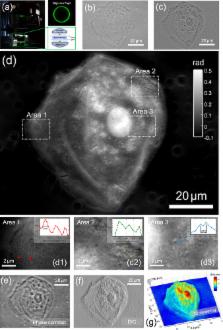
          For quantitative phase imaging (QPI) based on transport-of-intensity equation (TIE), partially coherent illumination provides speckle-free imaging, compatibility with brightfield microscopy, and transverse resolution beyond coherent diffraction limit. Unfortunately, in a conventional microscope with circular illumination aperture, partial coherence tends to diminish the phase contrast, exacerbating the inherent noise-to-resolution tradeoff in TIE imaging, resulting in strong low-frequency artifacts and compromised imaging resolution. Here, we demonstrate how these issues can be effectively addressed by replacing the conventional circular illumination aperture with an annular one. The matched annular illumination not only strongly boosts the phase contrast for low spatial frequencies, but significantly improves the practical imaging resolution to near the incoherent diffraction limit. By incorporating high-numerical aperture (NA) illumination as well as high-NA objective, it is shown, for the first time, that TIE phase imaging can achieve a transverse resolution up to 208 nm, corresponding to an effective NA of 2.66. Time-lapse imaging of in vitro Hela cells revealing cellular morphology and subcellular dynamics during cells mitosis and apoptosis is exemplified. Given its capability for high-resolution QPI as well as the compatibility with widely available brightfield microscopy hardware, the proposed approach is expected to be adopted by the wider biology and medicine community.

          Related collections

          Most cited references82

          • Record: found
          • Abstract: found
          • Article: not found

          Surpassing the lateral resolution limit by a factor of two using structured illumination microscopy.

          Lateral resolution that exceeds the classical diffraction limit by a factor of two is achieved by using spatially structured illumination in a wide-field fluorescence microscope. The sample is illuminated with a series of excitation light patterns, which cause normally inaccessible high-resolution information to be encoded into the observed image. The recorded images are linearly processed to extract the new information and produce a reconstruction with twice the normal resolution. Unlike confocal microscopy, the resolution improvement is achieved with no need to discard any of the emission light. The method produces images of strikingly increased clarity compared to both conventional and confocal microscopes.
            Bookmark
            • Record: found
            • Abstract: found
            • Article: not found

            Phase retrieval algorithms: a comparison

            J Fienup (1982)
            Iterative algorithms for phase retrieval from intensity data are compared to gradient search methods. Both the problem of phase retrieval from two intensity measurements (in electron microscopy or wave front sensing) and the problem of phase retrieval from a single intensity measurement plus a non-negativity constraint (in astronomy) are considered, with emphasis on the latter. It is shown that both the error-reduction algorithm for the problem of a single intensity measurement and the Gerchberg-Saxton algorithm for the problem of two intensity measurements converge. The error-reduction algorithm is also shown to be closely related to the steepest-descent method. Other algorithms, including the input-output algorithm and the conjugate-gradient method, are shown to converge in practice much faster than the error-reduction algorithm. Examples are shown.
              Bookmark
              • Record: found
              • Abstract: found
              • Article: not found

              Wide-field, high-resolution Fourier ptychographic microscopy

              In this article, we report an imaging method, termed Fourier ptychographic microscopy (FPM), which iteratively stitches together a number of variably illuminated, low-resolution intensity images in Fourier space to produce a wide-field, high-resolution complex sample image. By adopting a wavefront correction strategy, the FPM method can also correct for aberrations and digitally extend a microscope’s depth-of-focus beyond the physical limitations of its optics. As a demonstration, we built a microscope prototype with a resolution of 0.78 μm, a field-of-view of ~120 mm2, and a resolution-invariant depth-of-focus of 0.3 mm (characterized at 632 nm). Gigapixel colour images of histology slides verify FPM’s successful operation. The reported imaging procedure transforms the general challenge of high-throughput, high-resolution microscopy from one that is coupled to the physical limitations of the system’s optics to one that is solvable through computation.
                Bookmark

                Author and article information

                Contributors
                zuochao@njust.edu.cn
                chenqian@njust.edu.cn
                Journal
                Sci Rep
                Sci Rep
                Scientific Reports
                Nature Publishing Group UK (London )
                2045-2322
                9 August 2017
                9 August 2017
                2017
                : 7
                : 7654
                Affiliations
                [1 ]ISNI 0000 0000 9116 9901, GRID grid.410579.e, Smart Computational Imaging (SCI) Laboratory, , Nanjing University of Science and Technology, ; Nanjing, Jiangsu Province 210094 China
                [2 ]ISNI 0000 0000 9116 9901, GRID grid.410579.e, Jiangsu Key Laboratory of Spectral Imaging & Intelligent Sense, , Nanjing University of Science and Technology, ; Nanjing, Jiangsu Province 210094 China
                [3 ]ISNI 0000 0001 2224 0361, GRID grid.59025.3b, Centre for Optical and Laser Engineering (COLE), School of Mechanical and Aerospace Engineering, , Nanyang Technological University, ; Singapore, 639798 Singapore
                Author information
                http://orcid.org/0000-0002-1461-0032
                Article
                6837
                10.1038/s41598-017-06837-1
                5550517
                28794472
                6acda920-48b2-48dc-b1ac-7c19d2d02931
                © The Author(s) 2017

                Open Access This article is licensed under a Creative Commons Attribution 4.0 International License, which permits use, sharing, adaptation, distribution and reproduction in any medium or format, as long as you give appropriate credit to the original author(s) and the source, provide a link to the Creative Commons license, and indicate if changes were made. The images or other third party material in this article are included in the article’s Creative Commons license, unless indicated otherwise in a credit line to the material. If material is not included in the article’s Creative Commons license and your intended use is not permitted by statutory regulation or exceeds the permitted use, you will need to obtain permission directly from the copyright holder. To view a copy of this license, visit http://creativecommons.org/licenses/by/4.0/.

                History
                : 1 March 2017
                : 7 June 2017
                Categories
                Article
                Custom metadata
                © The Author(s) 2017

                Uncategorized
                Uncategorized

                Comments

                Comment on this article