4
views
0
recommends
+1 Recommend
0 collections
    0
    shares
      • Record: found
      • Abstract: found
      • Article: found
      Is Open Access

      MicroRNA-4476 promotes glioma progression through a miR-4476/APC/β-catenin/c-Jun positive feedback loop

      research-article

      Read this article at

      Bookmark
          There is no author summary for this article yet. Authors can add summaries to their articles on ScienceOpen to make them more accessible to a non-specialist audience.

          Abstract

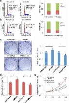
          Glioma has been a major healthcare burden; however, the specific molecular regulatory mechanism underlying its initiation and progression remains to be elucidated. Although it is known that many miRNAs are involved in the regulation of malignant phenotypes of glioma, the role of miR-4476 has not been reported yet. In the present study, we identify miR-4476 as an upregulated microRNA, which promotes cell proliferation, migration, and invasion in glioma. Further mechanistic analyses indicate that the adenomatous polyposis coli (APC), a negative regulator of the Wnt/β-catenin signaling pathway, is a direct target of miR-4476 and mediates the oncogenic effects of miR-4476 in glioma. C-Jun, a downstream effector of the Wnt/β-catenin signaling, is upregulated by miR-4476 overexpression. In turn, c-Jun could positively regulate miR-4476 expression by binding to the upstream of its transcription start site (TSS). Furthermore, in our clinical samples, increased miR-4476 is an unfavorable prognostic factor, and its expression positively correlates with c-Jun expression but negatively correlates with that of APC. In conclusion, our study demonstrates that miR-4476 acts as a tumor enhancer, directly targeting APC to stimulate its own expression and promoting the malignant phenotypes of glioma.

          Related collections

          Most cited references23

          • Record: found
          • Abstract: found
          • Article: not found

          APC mutations occur early during colorectal tumorigenesis.

          Human tumorigenesis is associated with the accumulation of mutations both in oncogenes and in tumour suppressor genes. But in no common adult cancer have the mutations that are critical in the early stages of the tumorigenic process been defined. We have attempted to determine if mutations of the APC gene play such a role in human colorectal tumours, which evolve from small benign tumours (adenomas) to larger malignant tumours (carcinomas) over the course of several decades. Here we report that sequence analysis of 41 colorectal tumours revealed that the majority of colorectal carcinomas (60%) and adenomas (63%) contained a mutated APC gene. Furthermore, the APC gene met two criteria of importance for tumour initiation. First, mutations of this gene were found in the earliest tumours that could be analysed, including adenomas as small as 0.5 cm in diameter. Second, the frequency of such mutations remained constant as tumours progressed from benign to malignant stages. These data provide strong evidence that mutations of the APC gene play a major role in the early development of colorectal neoplasms.
            Bookmark
            • Record: found
            • Abstract: found
            • Article: found
            Is Open Access

            MicroRNA-494 promotes cancer progression and targets adenomatous polyposis coli in colorectal cancer

            Background Aberrant activation of the Wnt/β-catenin signaling pathway is frequently observed in colorectal cancer (CRC). β-catenin is the major Wnt signaling pathway effector and inactivation of adenomatous polyposis coli (APC) results in nuclear accumulation of β-catenin. It has been suggested that inactivation of APC plays an important role in activation of the Wnt/β-catenin pathway and in the progression of colorectal tumorigenesis. However, the mechanism through which APC mediates colorectal tumorigenesis is not understood. Increasing evidence suggests that the dysregulation of microRNAs (miRNAs) is involved in colorectal tumorigenesis. Although miR-494 has been reported as being an upregulated miRNA, the interplay between miR-494 and APC-mediated colorectal tumorigenesis progression remains unclear. Methods The expression of miR-494 in tissues from patients diagnosed with CRC was analyzed using a microarray and real-time PCR. The effects of miR-494 on cell proliferation and tumorigenesis in CRC cells were analyzed by flow cytometry, colony formation assays, BrdU incorporation assays, and CCK8 assays. The correlation between miR-494 expression and APC expression, as well as the mechanisms by which miR-494 regulates APC in CRC were also addressed. Results miR-494 was significantly upregulated in CRC tissues, and this increase was negatively associated with APC expression. APC was confirmed to be a direct target of miR-494 in CRC. Furthermore, overexpression of miR-494 induced Wnt/β-catenin signaling by targeting APC, thus promoting CRC cell growth. Conclusions This study provides novel insights into the role of miR-494 in controlling CRC cell proliferation and tumorigenesis, and identifies miR-494 as a potential prognostic marker and therapeutic target.
              Bookmark
              • Record: found
              • Abstract: found
              • Article: not found

              Hepatitis C virus-induced up-regulation of microRNA-155 promotes hepatocarcinogenesis by activating Wnt signaling.

              Hepatitis C virus (HCV) infection usually induces chronic hepatic inflammation, which favors the initiation and progression of hepatocellular carcinoma (HCC). Moreover, microRNA-155 (miR-155) plays an important role in regulating both inflammation and tumorigenesis. However, little is known about whether and how miR-155 provides the link between inflammation and cancer. In this study we found that miR-155 levels were markedly increased in patients infected with HCV. MiR-155 transcription was regulated by nuclear factor kappa B (NF-κB), and p300 increased NF-κB-dependent miR-155 expression. The overexpression of miR-155 significantly inhibited hepatocyte apoptosis and promoted cell proliferation, whereas miR-155 inhibition induced G(0) /G(1) arrest. Up-regulated miR-155 resulted in nuclear accumulation of β-catenin and a concomitant increase in cyclin D1, c-myc, and survivin. Gain-of-function and loss-of-function studies demonstrated that miR-155 promoted hepatocyte proliferation and tumorigenesis by increasing Wnt signaling in vitro and in vivo, and DKK1 (Wnt pathway inhibitor) overexpression inhibited the biological role of miR-155 in hepatocytes. Finally, adenomatous polyposis coli (APC), which negatively regulates Wnt signaling, was identified as the direct and functional target of miR-155. HCV-induced miR-155 expression promotes hepatocyte proliferation and tumorigenesis by activating Wnt signaling. The present study provides a better understanding of the relationship between inflammation and tumorigenesis, and thus may be helpful in the development of effective diagnosis and treatment strategies against HCV-HCC. Copyright © 2012 American Association for the Study of Liver Diseases.
                Bookmark

                Author and article information

                Contributors
                xijiqi@126.com
                songye@smu.edu.cn
                zxa@smu.edu.cn
                Journal
                Cell Death Dis
                Cell Death Dis
                Cell Death & Disease
                Nature Publishing Group UK (London )
                2041-4889
                23 April 2020
                23 April 2020
                April 2020
                : 11
                : 4
                : 269
                Affiliations
                [1 ]GRID grid.416466.7, Department of Neurosurgery, , Nanfang Hospital, Southern Medical University, ; 510515 Guangzhou, Guangdong PR China
                [2 ]GRID grid.452437.3, Department of Neurosurgery, , The First Affiliated Hospital of Gannan Medical University, ; 341000 Ganzhou, Jiangxi PR China
                [3 ]ISNI 0000000123704535, GRID grid.24516.34, Department of Neurosurgery, Shanghai Tenth People’s Hospital, , Tongji University School of Medicine, ; 200072 Shanghai, PR China
                [4 ]Department of Neurosurgery, The First Hospital of Quanzhou Affiliated to Fujian Medical University, 362000 Quanzhou, Fujian Province PR China
                [5 ]Department of Neurosurgery, The First Hospital of Qiqihar City, 161005 Qiqihar, PR China
                Author information
                http://orcid.org/0000-0002-2641-1911
                Article
                2474
                10.1038/s41419-020-2474-4
                7181615
                32327666
                6a858bbf-2408-4a0e-98d0-a849960ecaf8
                © The Author(s) 2020

                Open Access This article is licensed under a Creative Commons Attribution 4.0 International License, which permits use, sharing, adaptation, distribution and reproduction in any medium or format, as long as you give appropriate credit to the original author(s) and the source, provide a link to the Creative Commons license, and indicate if changes were made. The images or other third party material in this article are included in the article’s Creative Commons license, unless indicated otherwise in a credit line to the material. If material is not included in the article’s Creative Commons license and your intended use is not permitted by statutory regulation or exceeds the permitted use, you will need to obtain permission directly from the copyright holder. To view a copy of this license, visit http://creativecommons.org/licenses/by/4.0/.

                History
                : 19 December 2019
                : 7 April 2020
                : 8 April 2020
                Funding
                Funded by: Startup Fund for Scientific Research of Fujian Medical University (Grant number;2018QH1186).
                Funded by: National Nature Science Fund of China (Grant NO. 81872064), Outstanding Youths Development Scheme of Nanfang Hospital, Southern Medical University (Grant NO. 2016J008), Natural Science Fund of Tibet Autonomous Region, China (Grant NO. XZ2017ZR-ZYZ27); Natural Science Foundation of Guangdong Province, China (Grant NO. 2020A151501122)
                Funded by: Science and Technology Program of Guangzhou, China (Grant NO. 201903010048)
                Categories
                Article
                Custom metadata
                © The Author(s) 2020

                Cell biology
                tumour biomarkers,mirnas,prognostic markers
                Cell biology
                tumour biomarkers, mirnas, prognostic markers

                Comments

                Comment on this article