27
views
0
recommends
+1 Recommend
0 collections
    0
    shares
      • Record: found
      • Abstract: found
      • Article: found
      Is Open Access

      Linc00152 promotes Cancer Cell Proliferation and Invasion and Predicts Poor Prognosis in Lung adenocarcinoma

      research-article

      Read this article at

      Bookmark
          There is no author summary for this article yet. Authors can add summaries to their articles on ScienceOpen to make them more accessible to a non-specialist audience.

          Abstract

          Background: The long non-coding RNA Linc00152 stimulates tumor progression in cancer. However, its clinical significance and biological functions in lung adenocarcinoma remains unknown. We evaluate the expression of Linc00152 in lung adenocarcinoma and its possible correlation with clinicopathologic features and patient survival to reveal its biological effects in cancer progression and prognosis.

          Methods: Total RNA extraction was performed on 110 pairs of lung adenocarcinoma and adjacent normal tissue samples, and then RT-qPCR was conducted. Chi-square test analysis was used to calculate the correlation between pathological parameters and the Linc00152 mRNA levels. Kaplan-Meier and Cox proportional hazards analyses were used to analyze the overall survival (OS) and disease-free survival (DFS) rates. We also detected the potential functional effects of overexpression and knockdown of Linc00152 in vitro cell proliferation, tumor cell invasion and migration, as well as in vivo nude mouse xenograft and metastasis models.

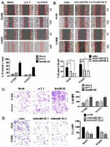
          Results: The Linc00152 expression levels were higher in lung adenocarcinoma samples than in the adjacent normal tissues. Linc00152 expression levels tightly correlated with lymph node metastasis station, remote metastasis and TNM staging. The Kaplan-Meier analysis suggested that high Linc00152 expression caused significantly poorer OS and DFS rates, and a multivariate analysis revealed that Linc00152 was an independent risk factor for both DFS and OS. Overexpression of Linc00152 in lung cancer cells stimulated proliferation, tumor cell invasion and migration. Knockdown of Linc00152 inhibited cell growth and cell invasion and migration. Finally, Linc00152 knockdown inhibited lung tumor growth and tumor metastasis in nude mice models.

          Conclusions: Our study suggests that Linc00152 independently predicts poor prognosis and promotes tumor progression in lung adenocarcinoma. Linc00152 needs to be considered as a potential molecular target in future cancer pharmacology.

          Related collections

          Most cited references16

          • Record: found
          • Abstract: found
          • Article: not found

          Long non-coding RNA metastasis associated in lung adenocarcinoma transcript 1 derived miniRNA as a novel plasma-based biomarker for diagnosing prostate cancer.

          Examining plasma RNA is an emerging non-invasive diagnosis technique. However, whether tumour-derived long non-coding RNAs (lncRNAs) in plasma can be used as a novel approach to detect human prostate cancer (PCa) has not yet been established. The study was divided into three parts: (1) the characteristics of PCa-related lncRNA fragments were systematically studied in the plasma or serum of 25 patients; (2) the source of the circulating lncRNA fragments was explored in vitro and in vivo; and (3) the diagnostic performance of metastasis associated in lung adenocarcinoma transcript 1 (MALAT-1) derived (MD) miniRNA was validated in an independent cohort of 192 patients. The expression levels of lncRNAs were measured by quantitative real time polymerase chain reaction (qRT-PCR). The MD-miniRNA copies were calculated using a standard curve in an area under the ROC curve (AUC)-receiver operating characteristic (ROC) analysis. Genome-wide profiling revealed that MALAT-1 and prostate cancer gene 3 (PCA3) are overexpressed in PCa tissues. Plasma lncRNAs probably exist in the form of fragments in a stable form. MD-miniRNA enters cell culture medium at measurable levels, and MD-miniRNA derived from human PCa xenografts actually enters the circulation in vivo and can be measured to distinguish xenografted mice from controls. In addition, plasma MD-miniRNA levels are significantly elevated in PCa patients compared to non-PCa patients (p<0.001). At a cut-off of 867.8 MD-miniRNA copies per microlitre of plasma, the sensitivity is 58.6%, 58.6% and 43.5% and the specificity is 84.8%, 84.8% and 81.6% for discriminating PCa from non-PCa, positive biopsy from negative biopsy and positive biopsy from negative biopsy, respectively. We conclude that MD-miniRNA can be used as a novel plasma-based biomarker for PCa detection and can improve diagnostic accuracy by predicting prostate biopsy outcomes. Further large-scale studies are needed to confirm our findings. Copyright © 2013 Elsevier Ltd. All rights reserved.
            Bookmark
            • Record: found
            • Abstract: found
            • Article: not found

            Long non-coding RNA Linc00152 is involved in cell cycle arrest, apoptosis, epithelial to mesenchymal transition, cell migration and invasion in gastric cancer.

            Gastric cancer remains a serious threat to public health with high incidence and mortality worldwide. Accumulating evidence demonstrates that long non-coding RNAs (lncRNAs) play important roles in regulating gene expression and are involved in various pathological processes, including gastric cancer. To investigate the possible role of dysregulated lncRNAs in gastric cancer development, we performed lncRNA microarray and identified 3141 significantly differentially expressed lncRNAs in gastric cancer tissues. Next, some of deregulated lncRNAs were validated among about 60 paired gastric cancer specimens such as Linc00261, DKFZP434K028, RPL34-AS1, H19, HOTAIR and Linc00152. Our results found that the decline of DKFZP434K028 and RPL34-AS1, and the increased expression of Linc00152 positively correlated with larger tumor size. The high expression levels of HOTAIR were associated with lymphatic metastasis and poor differentiation. Since the biological roles of Linc00152 are largely unknown in gastric cancer pathogenesis, we assessed its functions by silencing its up-regulation in gastric cancer cells. We found that Linc00152 knockdown could inhibit cell proliferation and colony formation, promote cell cycle arrest at G1 phase, trigger late apoptosis, reduce the epithelial to mesenchymal transition (EMT) program, and suppress cell migration and invasion. Taken together, we delineate the gastric cancer lncRNA signature and demonstrate the oncogenic functions of Linc00152. These findings may have implications for developing lncRNA-based biomarkers for diagnosis and therapeutics for gastric cancer.
              Bookmark
              • Record: found
              • Abstract: found
              • Article: not found

              HULC and Linc00152 Act as Novel Biomarkers in Predicting Diagnosis of Hepatocellular Carcinoma.

              The alterations of long non-coding RNAs (lncRNAs) are related to multiple diseases. They can be detected in plasma as biomarkers for the diagnosis of multiple diseases. In this study, we aimed to determine the expression of circulating lncRNAs in human, which may be promising biomarkers for the diagnosis of hepatocellular carcinoma (HCC).
                Bookmark

                Author and article information

                Journal
                J Cancer
                J Cancer
                jca
                Journal of Cancer
                Ivyspring International Publisher (Sydney )
                1837-9664
                2017
                5 July 2017
                : 8
                : 11
                : 2042-2050
                Affiliations
                [1 ]Department of Pathology, Fudan University Shanghai Cancer Center, Shanghai 200032, China
                [2 ]Department of Pathology, Obstetrics and Gynecology Hospital of Fudan University, Shanghai, 200011, China
                [3 ]Department of Respiratory Medicine, Shanghai Changzheng Hospital, Second Military Medical University, Shanghai 200003, China
                [4 ]Department of Pathology, Rui Jin Hospital, School of Medicine, Shanghai Jiao Tong University, Shanghai 20025, China
                Author notes
                ✉ Corresponding authors: Dr. Professor C-F Wang, Department of Pathology, Rui Jin Hospital, Shanghai Jiao Tong University School of Medicine, 197, Rui Jin Er Road, Shanghai 200025, China. E-mail: wang_chaofu@ 123456163.com ; Tel: +86-21-64175590; Fax: +86-21-64174774. And Dr. M-D Xu, Department of Pathology, Fudan University, Shanghai Cancer Center, 270 Dong'an Road, Shanghai 200032, China. Email: xumd27202003@ 123456sina.com ; Tel: +86-21-64175590.

                *Pei-pei Zhang, Yi-qin Wang and Wei-wei Weng shared the first-authorships.

                Competing Interests: The authors have declared that no competing interests exist.

                Article
                jcav08p2042
                10.7150/jca.18852
                5559966
                28819405
                69f8eb59-9ddc-40a7-8351-cf2fd276ab1c
                © Ivyspring International Publisher

                This is an open access article distributed under the terms of the Creative Commons Attribution (CC BY-NC) license ( https://creativecommons.org/licenses/by-nc/4.0/). See http://ivyspring.com/terms for full terms and conditions.

                History
                : 21 December 2016
                : 25 March 2017
                Categories
                Research Paper

                Oncology & Radiotherapy
                linc00152,lung adenocarcinoma,prognosis,proliferation,invasion
                Oncology & Radiotherapy
                linc00152, lung adenocarcinoma, prognosis, proliferation, invasion

                Comments

                Comment on this article

                scite_
                0
                0
                0
                0
                Smart Citations
                0
                0
                0
                0
                Citing PublicationsSupportingMentioningContrasting
                View Citations

                See how this article has been cited at scite.ai

                scite shows how a scientific paper has been cited by providing the context of the citation, a classification describing whether it supports, mentions, or contrasts the cited claim, and a label indicating in which section the citation was made.

                Similar content368

                Cited by22

                Most referenced authors244