14
views
0
recommends
+1 Recommend
0 collections
    0
    shares
      • Record: found
      • Abstract: found
      • Article: found
      Is Open Access

      Differentially expressed mRNAs, lncRNAs, and miRNAs with associated co-expression and ceRNA networks in ankylosing spondylitis

      research-article

      Read this article at

      Bookmark
          There is no author summary for this article yet. Authors can add summaries to their articles on ScienceOpen to make them more accessible to a non-specialist audience.

          Abstract

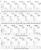
          Ankylosing spondylitis (AS) is a chronic autoimmune disease characterized by systemic inflammation and pathological osteogenesis. However, the genetic etiology of AS remains largely unknown. This study aimed to explore the potential role of coding and noncoding genes in the genetic mechanism of AS. Using microarray analyses, this study comprehensively compared lncRNA, microRNA, and mRNA profiles in hip joint ligament tissues from patients with AS and controls. A total of 661 lncRNAs, 574 mRNAs, and 22 microRNAs were differentially expressed in patients with AS compared with controls. Twenty-two of these genes were then validated using real-time polymerase chain reaction. Gene ontology and pathway analyses were performed to explore the principal functions of differentially expressed genes. The pathways were involved mainly in immune regulation, intercellular signaling, osteogenic differentiation, protein synthesis, and degradation. Gene signal transduction network, coding–noncoding co-expression network, and competing endogenous RNA expression network were constructed using bioinformatics methods. Then, two miRNAs, miR-17-5p and miR-27b-3p, that could increase the osteogenic differentiation potentials of ligament fibroblasts were identified. Finally, differentially expressed, five lncRNAs, four miRNAs, and five mRNAs were validated using quantitative real-time polymerase chain reaction. These results suggested that mRNAs, lncRNAs, and microRNAs were involved in AS pathogenesis. The findings might help characterize the pathogenesis of AS and provide novel therapeutic targets for patients with AS in the future.

          Related collections

          Most cited references41

          • Record: found
          • Abstract: not found
          • Article: not found

          Ankylosing Spondylitis and Axial Spondyloarthritis

            Bookmark
            • Record: found
            • Abstract: found
            • Article: found
            Is Open Access

            Plasma and synovial fluid microRNAs as potential biomarkers of rheumatoid arthritis and osteoarthritis

            Introduction MicroRNAs (miRNAs), endogenous small noncoding RNAs regulating the activities of target mRNAs and cellular processes, are present in human plasma in a stable form. In this study, we investigated whether miRNAs are also stably present in synovial fluids and whether plasma and synovial fluid miRNAs could be biomarkers of rheumatoid arthritis (RA) and osteoarthritis (OA). Methods We measured concentrations of miR-16, miR-132, miR-146a, miR-155 and miR-223 in synovial fluid from patients with RA and OA, and those in plasma from RA, OA and healthy controls (HCs) by quantitative reverse transcription-polymerase chain reaction. Furthermore, miRNAs in the conditioned medium of synovial tissues, monolayer fibroblast-like synoviocytes, and mononuclear cells were examined. Correlations between miRNAs and biomarkers or disease activities of RA were statistically examined. Results Synovial fluid miRNAs were present and as stable as plasma miRNAs for storage at -20°C and freeze-thawing from -20°C to 4°C. In RA and OA, synovial fluid concentrations of miR-16, miR-132, miR-146a, and miR-223 were significantly lower than their plasma concentrations, and there were no correlation between plasma and synovial fluid miRNAs. Interestingly, synovial tissues, fibroblast-like synoviocytes, and mononuclear cells secreted miRNAs in distinct patterns. The expression patterns of miRNAs in synovial fluid of OA were similar to miRNAs secreted by synovial tissues. Synovial fluid miRNAs of RA were likely to originate from synovial tissues and infiltrating cells. Plasma miR-132 of HC was significantly higher than that of RA or OA with high diagnosability. Synovial fluid concentrations of miR-16, miR-146a miR-155 and miR-223 of RA were significantly higher than those of OA. Plasma miRNAs or ratio of synovial fluid miRNAs to plasma miRNAs, including miR-16 and miR-146a, significantly correlated with tender joint counts and 28-joint Disease Activity Score. Conclusions Plasma miRNAs had distinct patterns from synovial fluid miRNAs, which appeared to originate from synovial tissue. Plasma miR-132 well differentiated HCs from patients with RA or OA, while synovial fluid miRNAs differentiated RA and OA. Furthermore, plasma miRNAs correlated with the disease activities of RA. Thus, synovial fluid and plasma miRNAs have potential as diagnostic biomarkers for RA and OA and as a tool for the analysis of their pathogenesis.
              Bookmark
              • Record: found
              • Abstract: found
              • Article: found
              Is Open Access

              Characterization of long non-coding RNA-associated ceRNA network to reveal potential prognostic lncRNA biomarkers in human ovarian cancer

              Accumulating evidence has underscored the important roles of long non-coding RNAs (lncRNAs) acting as competing endogenous RNAs (ceRNAs) in cancer initiation and progression. In this study, we used an integrative computational method to identify miRNA-mediated ceRNA crosstalk between lncRNAs and mRNAs, and constructed global and progression-related lncRNA-associated ceRNA networks (LCeNETs) in ovarian cancer (OvCa) based on “ceRNA hypothesis”. The constructed LCeNETs exhibited small world, modular architecture and high functional specificity for OvCa. Known OvCa-related genes tended to be hubs and occurred preferentially in the functional modules. Ten lncRNA ceRNAs were identified as potential candidates associated with stage progression in OvCa using ceRNA-network driven method. Finally, we developed a ten-lncRNA signature which classified patients into high- and low-risk subgroups with significantly different survival outcomes. Our study will provide novel insight for better understanding of ceRNA-mediated gene regulation in progression of OvCa and facilitate the identification of novel diagnostic and therapeutic lncRNA ceRNAs for OvCa.
                Bookmark

                Author and article information

                Journal
                Oncotarget
                Oncotarget
                Oncotarget
                ImpactJ
                Oncotarget
                Impact Journals LLC
                1949-2553
                26 December 2017
                27 November 2017
                : 8
                : 69
                : 113543-113557
                Affiliations
                1 Department of Orthopedics, Changhai Hospital, Second Military Medical University, Shanghai, China
                Author notes
                Correspondence to: Weidong Xu, xuwdshanghaichyy@ 123456126.com
                [*]

                These authors contributed equally to this work

                Article
                22708
                10.18632/oncotarget.22708
                5768345
                29371928
                69ea0fb3-2c15-4d49-8dba-f6ac3b0a7a0b
                Copyright: © 2017 Zhang et al.

                This is an open-access article distributed under the terms of the Creative Commons Attribution License 3.0 (CC BY 3.0), which permits unrestricted use, distribution, and reproduction in any medium, provided the original author and source are credited.

                History
                : 21 September 2017
                : 29 October 2017
                Categories
                Research Paper

                Oncology & Radiotherapy
                ankylosing spondylitis,microarray,competing endogenous rna,long noncoding rna,mirna

                Comments

                Comment on this article