0
views
0
recommends
+1 Recommend
0 collections
    0
    shares
      • Record: found
      • Abstract: found
      • Article: found
      Is Open Access

      Magnetic Resonance Imaging in Cervical Spine Trauma: More Than Soft Tissue Illustration

      case-report
      1 , , 1 , 1
      ,
      Cureus
      Cureus
      computed tomography, misdiagnosis, magnetic resonance imaging, trauma, cervical-spine

      Read this article at

      Bookmark
          There is no author summary for this article yet. Authors can add summaries to their articles on ScienceOpen to make them more accessible to a non-specialist audience.

          Abstract

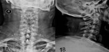
          The role of magnetic resonance imaging (MRI) in cervical spine trauma is limited to visualizing soft tissues such as the intervertebral disc, the spinal cord, and hematomas. Herein, we present the case of a 60-year-old man who was transferred to our hospital with neck pain after a cervical spine trauma associated with a motor vehicle accident. The initial computed tomography imaging of the cervical spine showed stable linear fractures at the C2, C6, and C7 vertebral bodies, for which the patient received conservative management. The patient showed remarkable clinical improvement three months later, but the linear fractures at the subaxial spine remained unchanged on computed tomography (CT). Magnetic resonance imaging (MRI) scantly differentiated active from inactive bone lesions and prevented unnecessary interventions. Therefore, we suggest that the MRI is of value in cases with a clinical and radiological mismatch. A mismatch is considered in cases when there is a high level of clinical suspicion for a spinal fracture, whereas CT images fail to provide direct evidence of a bone fracture. In such cases, MRI offers indirect evidence of bony trauma, such as bone marrow edema, visualized as a high-intensity signal in T2-weighted images. Furthermore, specialized spine trauma MRI protocols could be of value in selected cases.

          Related collections

          Most cited references7

          • Record: found
          • Abstract: found
          • Article: not found

          BLADE in sagittal T2-weighted MR imaging of the cervical spine.

          Image quality and diagnostic reliability of T2-weighted MR images of the cervical spine are often impaired by several kinds of artifacts, even in cooperative patients. The aim of this study was to evaluate if BLADE sequences might solve these problems in a routine patient collective.
            Bookmark
            • Record: found
            • Abstract: found
            • Article: not found

            Utility of MRI for cervical spine clearance in blunt trauma patients after a negative CT.

            To determine the utility of cervical spine MRI in blunt trauma evaluation for instability after a negative non-contrast cervical spine CT.
              Bookmark
              • Record: found
              • Abstract: found
              • Article: found
              Is Open Access

              Cervical Spine Computed Tomography Imaging Artifact Affecting Clinical Decision-Making in the Traumatized Patient

              CT scanning is an important tool in the evaluation of trauma patients. We review a case involving a trauma patient in which a cervical spine computed tomography (CT) artifact affected decision-making by physicians. The CT artifact mimicked bilateral dislocated facets (51-B1.1). On the basis of CT findings, the patient was transferred to a different hospital for evaluation. Discrepancy between the primary CT scan and patient physical exam prompted secondary CT scans and X-ray evaluation; neither of these studies showed osseous abnormalities. This case reinforces the necessity for physicians to formulate their diagnosis based upon multiple areas of information including physical examination, plain x-ray and subsequent advanced imaging, rather than relying solely on advanced imaging.
                Bookmark

                Author and article information

                Journal
                Cureus
                Cureus
                2168-8184
                Cureus
                Cureus (Palo Alto (CA) )
                2168-8184
                22 January 2022
                January 2022
                : 14
                : 1
                : e21493
                Affiliations
                [1 ] Neurosurgery, General University Hospital of Larisa, Larisa, GRC
                Author notes
                George Fotakopoulos gfotakop@ 123456yahoo.gr
                Article
                10.7759/cureus.21493
                8860237
                694fb857-0308-419b-94da-f3f022f95159
                Copyright © 2022, Fotakopoulos et al.

                This is an open access article distributed under the terms of the Creative Commons Attribution License, which permits unrestricted use, distribution, and reproduction in any medium, provided the original author and source are credited.

                History
                : 21 January 2022
                Categories
                Neurology
                Radiology
                Neurosurgery

                computed tomography,misdiagnosis,magnetic resonance imaging,trauma,cervical-spine

                Comments

                Comment on this article