9
views
0
recommends
+1 Recommend
0 collections
    0
    shares
      • Record: found
      • Abstract: found
      • Article: found
      Is Open Access

      Protein and lipid mass concentration measurement in tissues by stimulated Raman scattering microscopy

      research-article

      Read this article at

      Bookmark
          There is no author summary for this article yet. Authors can add summaries to their articles on ScienceOpen to make them more accessible to a non-specialist audience.

          Significance

          We report a quantitative Raman microscopy method that measures the concentration of protein and lipid in cells at high spatial resolution in living and in fixed samples of tissues, allowing quantitative studies of cell size and organelle regulation both in cell culture and in tissue slices; it can be applied to problems of cell size control, intracellular crowding, and lipid metabolism in the context of cell growth, cell differentiation, cell senescence, and pathology.

          Abstract

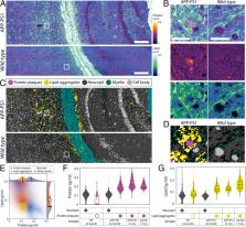
          Cell mass and chemical composition are important aggregate cellular properties that are especially relevant to physiological processes, such as growth control and tissue homeostasis. Despite their importance, it has been difficult to measure these features quantitatively at the individual cell level in intact tissue. Here, we introduce normalized Raman imaging (NoRI), a stimulated Raman scattering (SRS) microscopy method that provides the local concentrations of protein, lipid, and water from live or fixed tissue samples with high spatial resolution. Using NoRI, we demonstrate that protein, lipid, and water concentrations at the single cell are maintained in a tight range in cells under the same physiological conditions and are altered in different physiological states, such as cell cycle stages, attachment to substrates of different stiffness, or by entering senescence. In animal tissues, protein and lipid concentration varies with cell types, yet an unexpected cell-to-cell heterogeneity was found in cerebellar Purkinje cells. The protein and lipid concentration profile provides means to quantitatively compare disease-related pathology, as demonstrated using models of Alzheimer’s disease. This demonstration shows that NoRI is a broadly applicable technique for probing the biological regulation of protein mass, lipid mass, and water mass for studies of cellular and tissue growth, homeostasis, and disease.

          Related collections

          Most cited references71

          • Record: found
          • Abstract: not found
          • Article: not found

          Hallmarks of Cellular Senescence

            Bookmark
            • Record: found
            • Abstract: found
            • Article: not found

            Trainable Weka Segmentation: a machine learning tool for microscopy pixel classification.

            State-of-the-art light and electron microscopes are capable of acquiring large image datasets, but quantitatively evaluating the data often involves manually annotating structures of interest. This process is time-consuming and often a major bottleneck in the evaluation pipeline. To overcome this problem, we have introduced the Trainable Weka Segmentation (TWS), a machine learning tool that leverages a limited number of manual annotations in order to train a classifier and segment the remaining data automatically. In addition, TWS can provide unsupervised segmentation learning schemes (clustering) and can be customized to employ user-designed image features or classifiers.
              Bookmark
              • Record: found
              • Abstract: found
              • Article: not found

              Label-free biomedical imaging with high sensitivity by stimulated Raman scattering microscopy.

              Label-free chemical contrast is highly desirable in biomedical imaging. Spontaneous Raman microscopy provides specific vibrational signatures of chemical bonds, but is often hindered by low sensitivity. Here we report a three-dimensional multiphoton vibrational imaging technique based on stimulated Raman scattering (SRS). The sensitivity of SRS imaging is significantly greater than that of spontaneous Raman microscopy, which is achieved by implementing high-frequency (megahertz) phase-sensitive detection. SRS microscopy has a major advantage over previous coherent Raman techniques in that it offers background-free and readily interpretable chemical contrast. We show a variety of biomedical applications, such as differentiating distributions of omega-3 fatty acids and saturated lipids in living cells, imaging of brain and skin tissues based on intrinsic lipid contrast, and monitoring drug delivery through the epidermis.
                Bookmark

                Author and article information

                Journal
                Proc Natl Acad Sci U S A
                Proc Natl Acad Sci U S A
                pnas
                PNAS
                Proceedings of the National Academy of Sciences of the United States of America
                National Academy of Sciences
                0027-8424
                1091-6490
                22 April 2022
                26 April 2022
                22 October 2022
                : 119
                : 17
                : e2117938119
                Affiliations
                [1] aDepartment of Systems Biology, Harvard Medical School , Boston, MA 02115;
                [2] bDepartment of Genetics, Harvard Medical School , Boston, MA 02115;
                [3] cCenter for Advanced Imaging, Harvard University , Cambridge, MA 20138;
                [4] dAthinoula A. Martinos Center for Biomedical Imaging, Massachusetts General Hospital and Harvard Medical School , Boston, MA 02129;
                [5] eDepartment of Chemistry, University of Washington , Seattle, WA 98195;
                [6] fBiomedical Pioneering Innovation Center, Peking University , Beijing 100871; China
                Author notes
                2To whom correspondence may be addressed. Email: danfu@ 123456uw.edu or marc@ 123456hms.harvard.edu .

                Contributed by Marc W. Kirschner; received October 6, 2021; accepted February 21, 2022; reviewed by Jennifer Lippincott-Schwartz, Matthieu Piel, Hervé Rigneault, and Lu Wei

                Author contributions: S.O., C.L., A.M., M.B., and M.W.K. designed research; S.O., C.L., A.M., and D.F. performed research; A.L., C.R., W. Yin, and D.F. contributed new reagents/tools; S.O., C.L., W. Yang, C.R., W. Yin, C.J.T., and X.S.X. analyzed data; S.O., C.L., and M.W.K. wrote the paper; and W. Yang and A.L. provided technical advice and manuscript revision.

                1S.O. and C.L. contributed equally to this work.

                Author information
                https://orcid.org/0000-0002-4064-9301
                https://orcid.org/0000-0002-3892-4108
                https://orcid.org/0000-0001-9243-8306
                https://orcid.org/0000-0001-6540-6130
                Article
                202117938
                10.1073/pnas.2117938119
                9169924
                35452314
                6925e66d-fd44-42c8-bbd1-4fb54cd98ee5
                Copyright © 2022 the Author(s). Published by PNAS.

                This article is distributed under Creative Commons Attribution-NonCommercial-NoDerivatives License 4.0 (CC BY-NC-ND).

                History
                : 21 February 2022
                Page count
                Pages: 11
                Funding
                Funded by: HHS | NIH | National Institute of General Medical Sciences (NIGMS) 100000057
                Award ID: R01GM026875
                Award Recipient : Markus Thomas Basan Award Recipient : Marc W. Kirschner
                Funded by: HHS | NIH | Eunice Kennedy Shriver National Institute of Child Health and Human Development (NICHD) 100009633
                Award ID: HD03443
                Award Recipient : ChangHee Lee Award Recipient : Clifford J. Tabin
                Funded by: HHS | NIH | National Institute of Biomedical Imaging and Bioengineering (NIBIB) 100000070
                Award ID: R01EB017254
                Award Recipient : Wenlong Yang Award Recipient : Ang Li Award Recipient : Xiaoliang Sunney Xie
                Funded by: HHS | NIH | National Institute on Aging (NIA) 100000049
                Award ID: R01AG055413
                Award Recipient : Chongzhao Ran Award Recipient : Wei Yin
                Funded by: HHS | NIH | National Institute of General Medical Sciences (NIGMS) 100000057
                Award ID: R35GM137895
                Award Recipient : Markus Thomas Basan Award Recipient : Marc W. Kirschner
                Categories
                408
                Biological Sciences
                Biophysics and Computational Biology

                cell size,quantitative microscopy,single cell mass quantification,lipid imaging,stimulated raman scattering

                Comments

                Comment on this article